Feline portosystemic shunt morphology
This is just a slightly unusual shunt conformation in a six-month old kitten.
As is common, the anomalous vessel originates from the base of the splenic vein and courses cranially and dorsally. The slightly unusual thing is that the confluence with the cava is intra-hepatic.
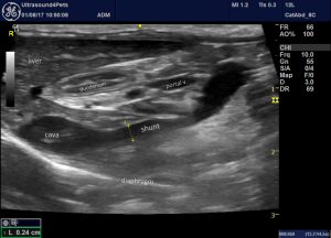
Longitudinal plane view from R cranioventral abdominal wall
With colour Doppler (clip starts with cava (above) and aorta (below), later angle shifts to show the shunt entering the cava and curving around from the caudal end of the portal vein):
And at surgery:
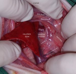
View at laparotomy: the duodenum has been reflected to the left to expose the omental foramen. Although the shunt is on the left of midline it can be accessed nicely through the foramen which avoids the need to open the omental bursa for a left-sided approach.
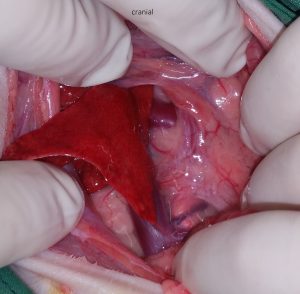
Without annotations:
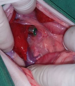
And with ameroid constrictor: