Feline hypoadrenocorticism
This is an 11 year-old female DSH with a history of progressive vague malaise, inappetence, weight loss and muscle weakness.
On physical examination, striking features were profound muscle weakness, skin tenting and non-palpable femoral pulses. She was not bradycardic.
Blood biochemistry:
urea 13.9 mmol/l (ref 3-10)
creatinine 210 umol/l (ref <180)
total calcium 3.17 (ref 2.0-3.0)
phosphorus 2.33 mmol/l (ref <1.6)
sodium 128 mmol/l (ref 145-156)
potassium 5.9 mmol/l (ref 4.0-5.0)
Ultrasonographic findings are not easy to convey with still images. My overwhelming impression at the time was that the whole abdominal exam was very difficult because all major vascular landmarks were hard to make out since they collapsed when any pressure was applied. This is the portal vein:
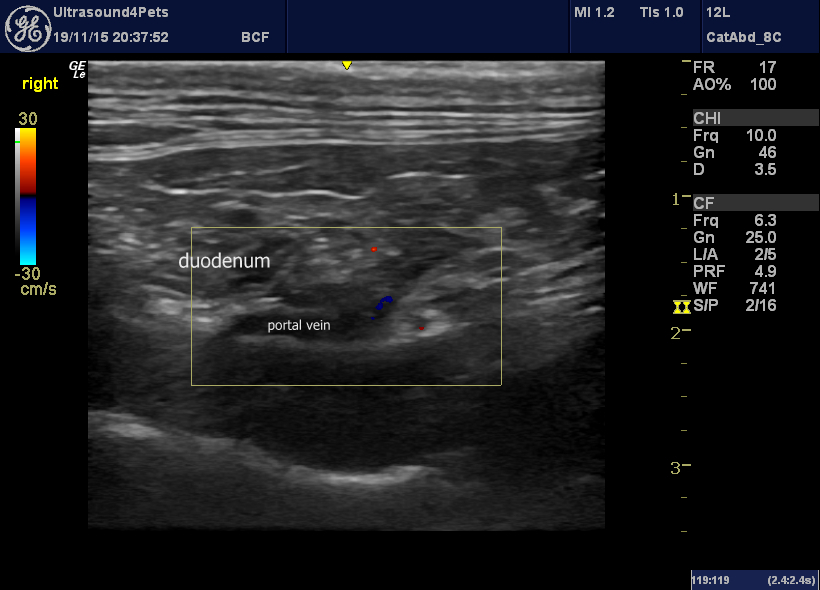
Longitudinal view of the porta hepatis from the right side showing a narrow portal vein
In this long-axis view of the heart the right atrium is also subjectively very small -indicative of reduced venous return.
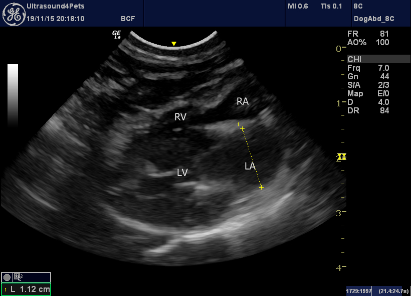
Right long-axis four-chamber view of the heart. The right atrium is small.
The kidneys appeared completely unremarkable although at the low end of normal length.
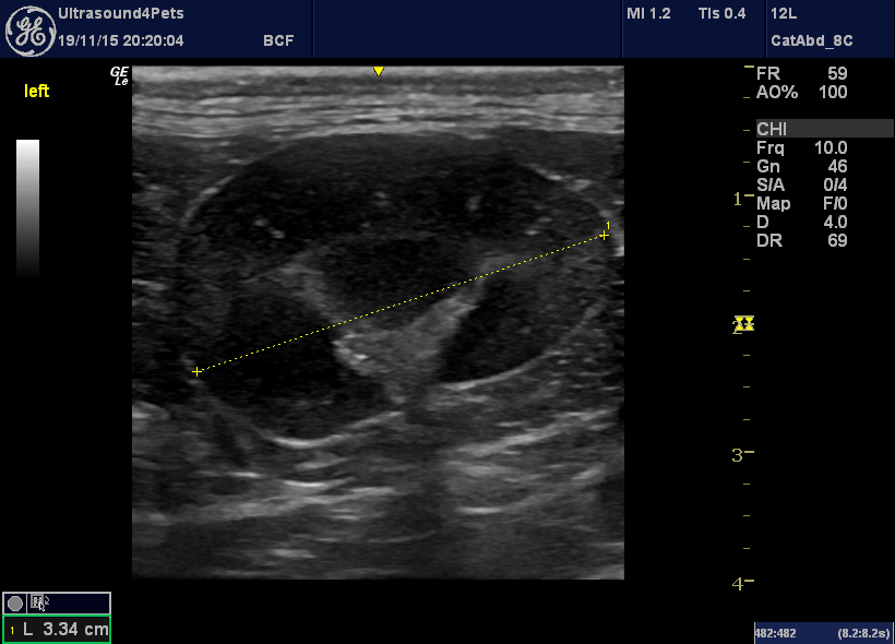
longitudinal view of the left kidney
The left adrenal was never convincingly identified and the right was very thin at 2.0mm diameter.
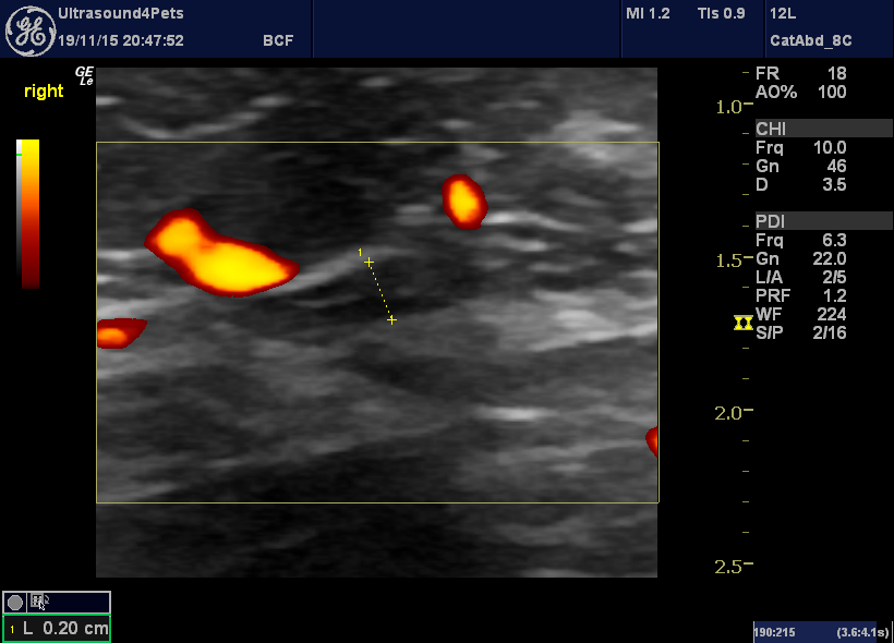
longitudinal plane view of the right adrenal
Blood pressure was measured at 75/35.
Blood cortisol remained unmeasurably low pre- and post-ACTH.


