Emphysematous cholecystitis in an elderly Border Terrier
This is just one of those things that tells you that someone up there has a sense of humour. The day after the aforementioned dog with emphysematous cystitis I was asked to look at a somewhat elderly Border Terrier with a history of well-controlled pituitary-dependent hyperadrencorticism but now presenting with acute abdominal pain and collapse.
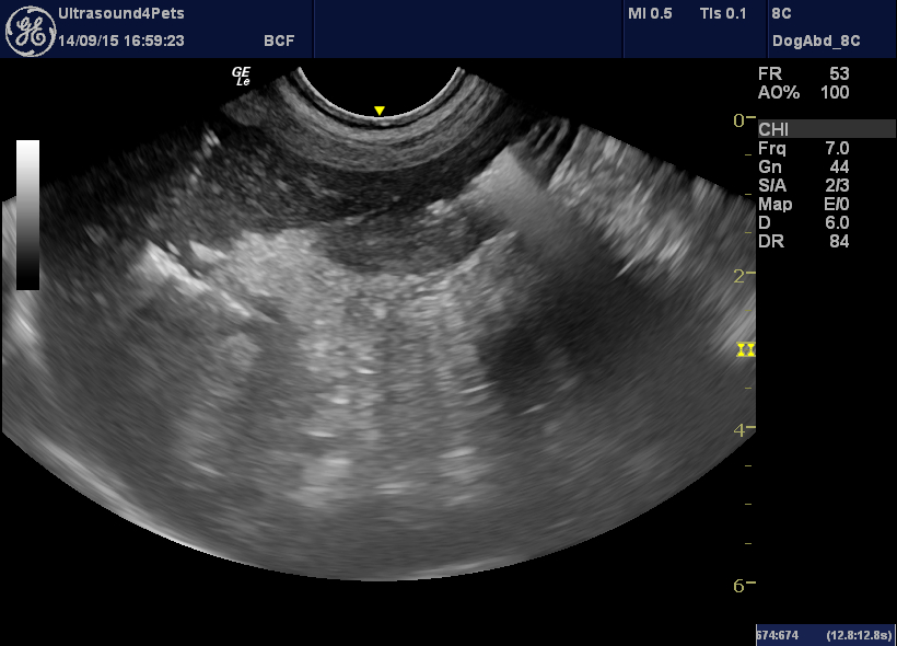
So, starting from the beginning, that cranial abdominal fat looks a little hyperechoic compared to normal -certainly quite a strong contrast with the hypoechoic liver in this image. But no effusion.
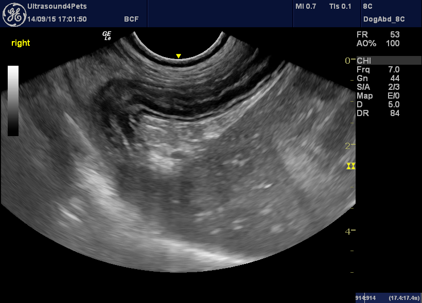
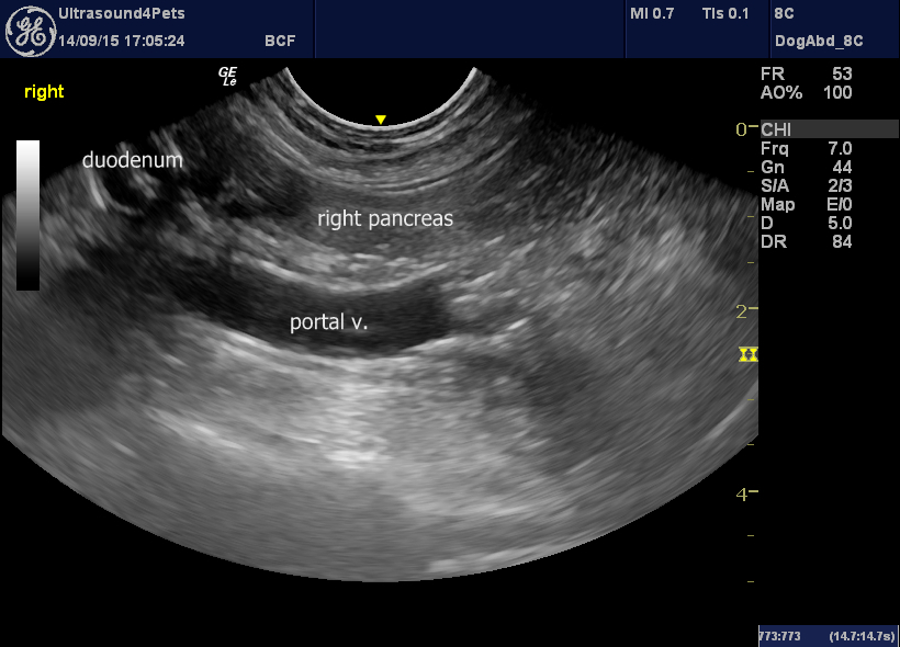
longitudinal plane view from the right cranial abdominal wall showing the cranial duodenal flexure, first part of descending duodenum and part of body of pancreas
It’s subtle but the duodenum is slightly corrugated. Not pronounced folding -as in the plication seen with linear foreign bodies- but a slight crinkliness of the walls. This is non-specific but also suggests an inflamed abdomen -as in pancreatitis, severe enteritis, septic peritonitis and so forth.
The pancreas…..
Nothing very exciting there. Certainly the right lobe and the body (previous image in the curve of the cranial duodenal flexure) have no convincing swelling or hypoechoic change. The hyperechoic abdominal fat is not focussed on the pancreas.
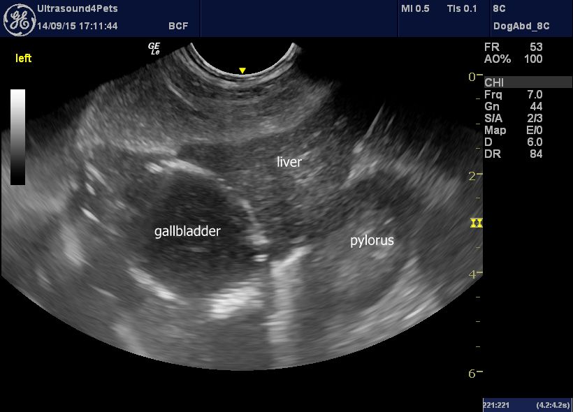
The diagnostic changes are in the first few views of the cranial abdomen:
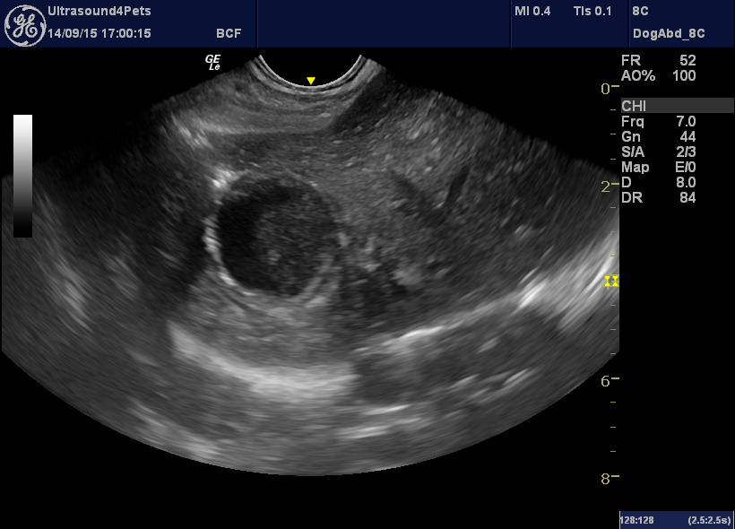
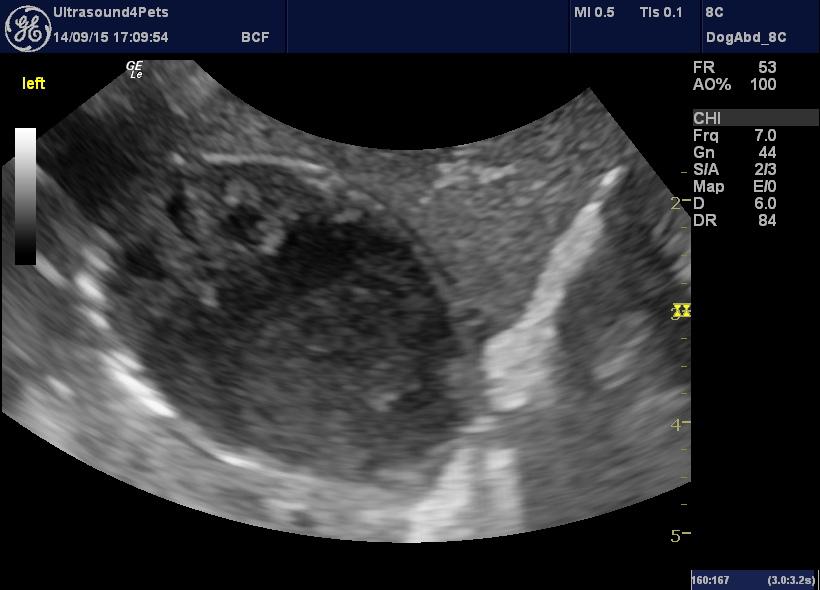
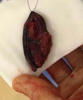
The gallbladder is large. But more importantly, there are distinct intensely-hyperechoic gas shadows around the outer margin of the wall. This is a gas-producing, emphysematous cholecystitis.
There is no evidence of hypoechoic mucus in the gallbladder so I presume that this is a septic cholangitis with HAC as a predisposing factor.
Having debated the matter, consulted the literature and concluded that there’s not much data out there we elected for surgery.
…..and, with the help of some metronidazole and co-amoxyclav, he’s never looked back.


