Double caudal vena cava, circumcaval ureter and ureteral calculus in a cat
There are just some practices where every second case is really unusual…..
This is a cat whose immediate problem is azotaemia due to a ureteral calculus (function of the other kidney is impaired -likely due to previous episodes of ureteral obstruction)
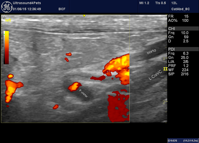
The image on the left is a frontal plane view from the left flank (with power Doppler to demonstrate blood vessels). The dilated right ureter can be seen looping around the the left of the caudal vena cava (circumcaval ureter). It is dilated to about 2mm (normal being @0.4mm).
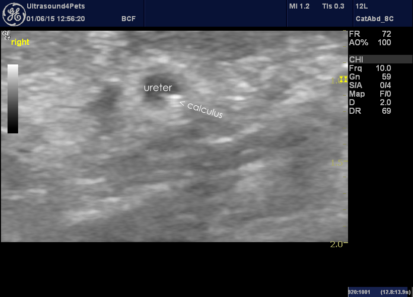
On the right is a frontal plane view from the right flank showing the termination of the dilated section of right ureter at an echogenic object which is presumably a calculus.
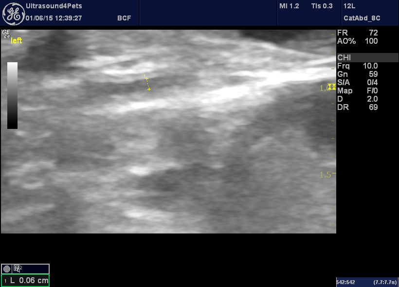
The left ureter is more or less normal -this degree of mild dilation is consistent with intravenous fluid therapy.
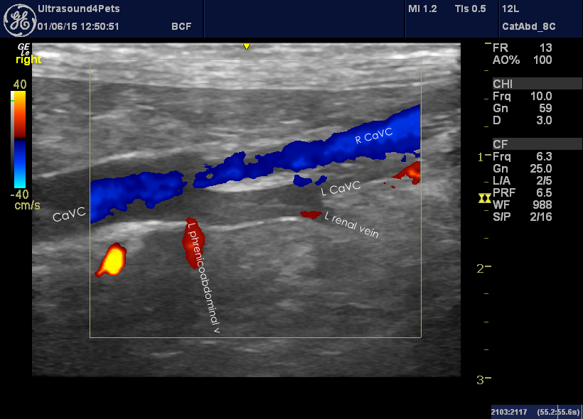
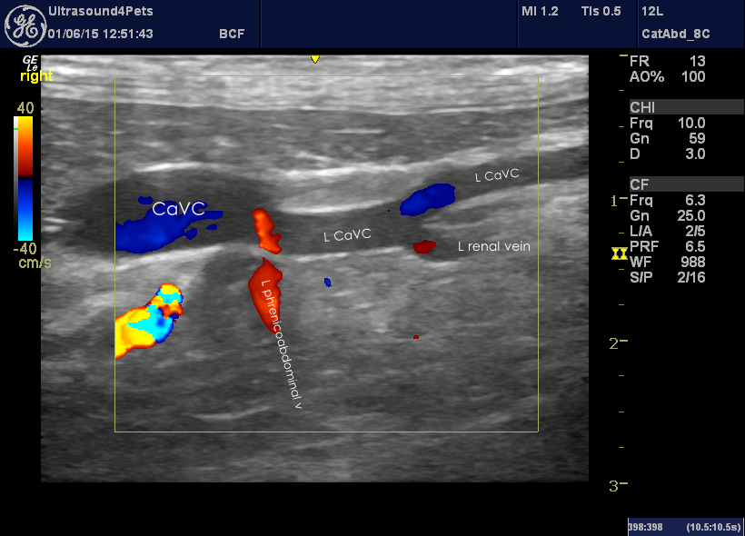
However, there is more going on here. A frontal plane view from the right showing the major vessels at mid-abdominal level is interesting:
There is a double caudal vena cava with the confluence just caudal to the left phrenicoabdominal vein. The left renal vein drains into the left caudal vena cava.
There is some evidence that this variant is not all that uncommon -Belanger and otjhers (Am J Vet Res Jan 2014) found that about 7% of 300 cats exhibited double CaVC.
Circumcaval right ureter is also not uncommon in cats. There has been some debate about the possibility that these cats might be at increased risk of ureteral obstruction at this ‘pinch point’ where the ureter wraps around the cava. And it’s tempting to believe it when you see the images. However, the one good review of this issue (Steinhaus et al. JVIM Jan 2015) found that the incidence of ureteral obstruction was the same in both normal and circumcaval arrangements.


