Delayed sonographic changes in acute feline pancreatitis
Recent research has documented delayed onset of sonographic changes in acute canine pancreatitis. This is enormously important in a clinical setting where we now realise that the absence of typical pancreatitis findings on first presentation doesn’t rule it out and we are sometimes going to have to re-scan a dog with ‘acute abdomen’ a few days down the line to get a full picture.
We’ve previously noted that the same happens in cats. This is a recent patient presented with acute onset depression, ptyalism, vomiting, complete anorexia and abdominal pain:
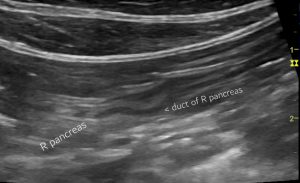
Day 2 after initial presentation: right sided longitudinal plane of the right lobe of pancreas. On full thoraco-abdominal sonographic examination there was no clear evidence of the cause of signs.
By day 5 however…
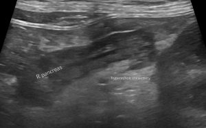
Same view day 5: now that looks like a convincing pancreatitis. There is now swelling, heteroechoic pancreatic parenchyma and markedly hyperechoic peripancreatic mesentery. This hyperechoic change is specifically focused on the pancreas on both left and right.
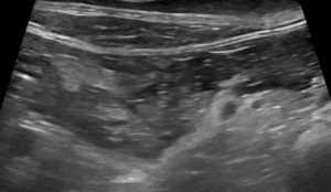
Day 5: slightly different plane of view, right pancreas


What paper is documenting the delayed sonographic changes in canine pancreatitis?
J Vet Intern Med 2022 May;36(3):947-956Prevalence of ultrasonographic gastrointestinal wall changes in dogs with acute pancreatitis: A retrospective study (2012-2020)Joshua J Hardwick, Elizabeth J Reeve, Melanie J Hezzell, J
the link is in the blog post as well Sarah