Congenital portosystemic shunt with left colic to caval shunting
This is a 5 month-old cat with recurrent haematuria.
The bladder:
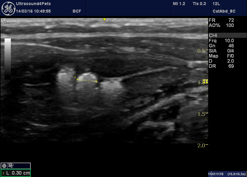
The bladder is almost empty apart from a collection of calculi up to 4mm diameter. And this is very suspicious in a young cat.
The vessels at the porta hepatis are interesting, although the portal vein and its tributaries are arranged normally the whole portal circulation in this area is narrower than normal. Furthermore the hepatic artery as it crosses the portal vein is large (red flow in centre of image).
That’s another pointer towards a portosystemic shunt. Normally the liver receives some 80% of its supply from the portal vein. In the presence of a shunt much of the portal flow is diverted directly into the systemic circulation. Compensatory hypertrophy of the hepatic arterial supply is usual.
Actually there is one abnormality in portal vein tributaries in this cat: the splenic vein should enter the portal vein as shown here:
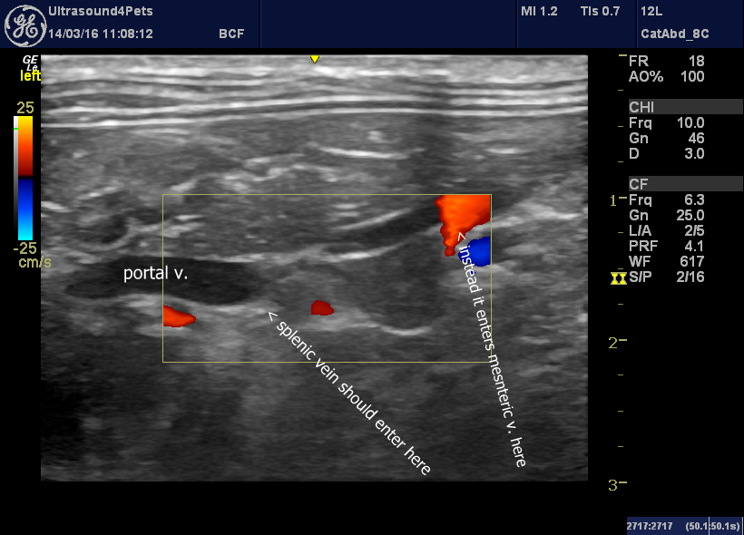
Instead it joins the mesenteric vein more caudally and from that point flow in the mesenteric vein is reversed – being directed away from the liver (hepatofugally)
The mesenteric vein can be followed caudally to mid abdomen where the cranial and caudal mesenteric veins meet:
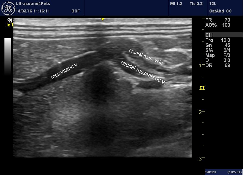
But, look at the colour Doppler:
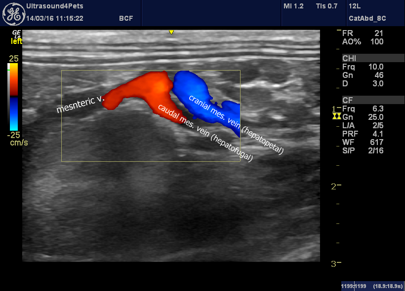
Whilst flow in the cranial mesenteric vein is hepatopetal, that in the caudal mesenteric continues caudally way from the liver.
Eventually, it loops to the left running as far as the aortic bifurcation and then turns cranially before emptying into the vena cava. This cat has a double vena cava at this level (not such an uncommon phenomenon). The shunt empties into the left cava.
This video clip follows the shunt in reverse from the cava as it loops round and then runs cranially:
And there is a large eccentric jet of mitral regurgitation probably due to congenital dysplasia.
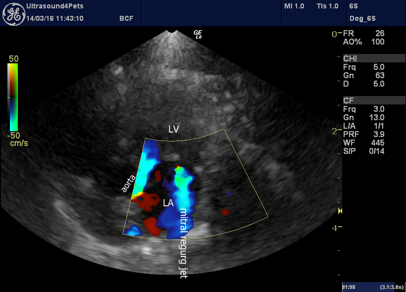
left apical view of the heart optimised for the mitral valve: there is a large regurgitant jet


