Congenital interruption of the portal vein in dogs: extrahepatic portosystemic shunts which are inoperable
In the vast majority of congenital extrahepatic shunts, the portal vein (PV) exists in normal relation to its tributaries and branches but is hypoplastic due to diversion of flow. Just occasionally, one comes across a shunt with complete absence or interruption of the PV.
Vet Surg. 1998 May-Jun;27(3):203-15.
Congenital interruption of the portal vein and caudal vena cava in dogs: six case reports and a review of the literature.
Hunt GB1, Bellenger CR, Borg R, Youmans KR, Tisdall PL, Malik R.
https://www.ncbi.nlm.nih.gov/pubmed/9605232
This is important because it’s not a good idea to occlude the shunt in this scenario! In the report cited above, 7% of dogs presented to the University of Sydney with portosystemic shunts during the study period exhibited interrupted PV: a surprisingly large proportion.
Strictly speaking, interruption should be proven with angiographic studies involving temporary occlusion of the shunt to try to force flow through the PV.
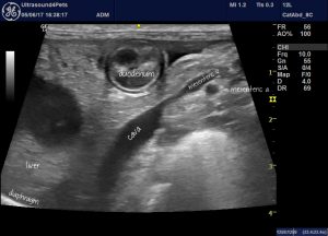
This is an example of such a case suspected on the basis of ultrasonographic findings:

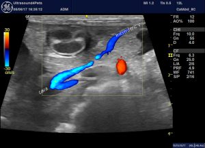
Longitudinal plane view from the right side showing the cava and its relation to the mesenteric vein. The latter empties completely into the former with no apparent portal vein present.
and with colour:

That’s also a very usual shunt configuration. Most congenital portosystemic shunts terminating in the cava do so via an anomalous left gastric vein entering on the left aspect cranial to the R adrenal and phrenicoabdominal vein.
As video:


