Congenital extrahepatic portosystemic shunt via the right gastric vein in a dog
Congenital extra-hepatic shunts in dogs most commonly involve diversion of blood from the portal system via either 1) the splenic vein and thence left gastric vein to the systemic circulation (cava, azygous or left phrenic veins) or 2) via the gastroduodenal vein and then right gastric vein to any of the same systemic veins.
This is just a particularly convincing example of a right gastric shunt.
In black/white views the caudal tributaries of the portal vein (mesenteric and splenic veins) appear in normal proportions. However, the gastroduodenal vein is grossly dilated whereas the continuation of the portal vein towards the liver is abnormally narrow:
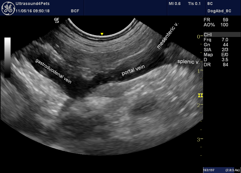
longitudinal plane view from a right paracostal view-point showing the portal vein and its tributaries
With colour, normal hepatopetal flow in the mesenteric and splenic veins is confirmed. But the gastroduodenal carries reversed, hepatofugal flow:
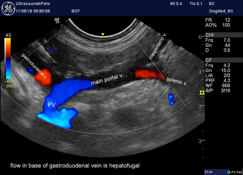
Same view with colour Doppler. The apparent continuity between the cranial part of the portal vein and the underlying cava is an artefact due to colour ‘bloom’.
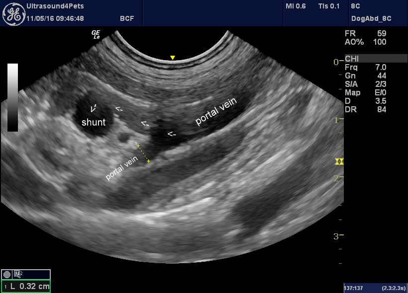
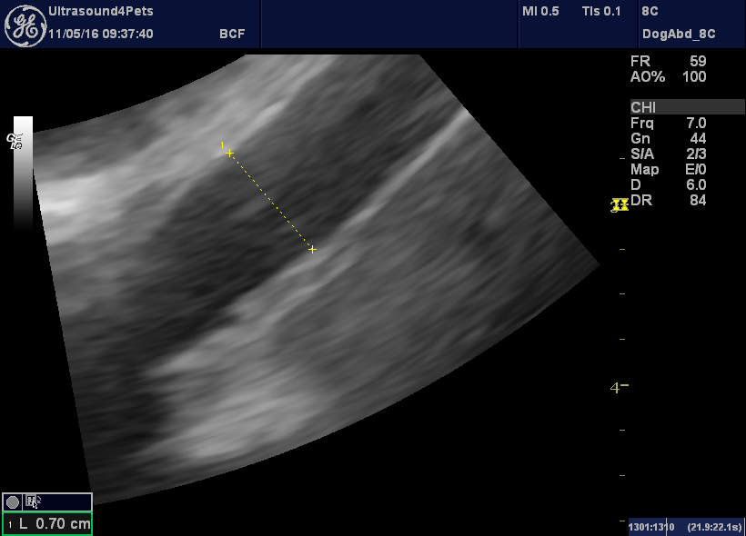
Similar view at slightly different angle showing continuation of the shunt into the right gastric vein and measurement of the portal vein cranial to the shunt origin.
This is where it gets more complicated. These right-sided shunts typically follow a looping course along the lesser curvature of the stomach and then join the cava on its left lateral aspect.
A great paper by White and Parry with nice CT angiographic images shows that the vessel entering the cava is actually an anomalous left gastric vein. This would normally join the splenic vein and thence the portal vein.
http://www.ncbi.nlm.nih.gov/pubmed/25871881
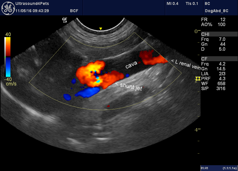
longitudinal plane view of the vena cava from a right paracostal viewpoint showing flow entering from the anomalous left gastric vein.
We don’t really need it here but the ratio of the portal vein diameter (cranial to the gastroduodenal vein) to aortic diameter is 3.2:7.0 = 0.45. Anything below 0.65 is strongly indicative of an extrahepatic shunt.
aortic diameter as seen from a right paracostal viewpoint


