Colonic torsion in a dog: ultrasonographic findings
The unfortunate patient in this case is a 4 y.o. FN German Shepherd. She has a history of chronic gastrointestinal disease. Previous sonography demonstrated severely thickened ileum and colon. TLI was subnormal. Full-thickness gut biopsies yielded a diagnosis of chronic enterocolitis. Subsequently, 6 months prior to the torsion, an apparent ileal impaction necessitated resection of the ileo-caeco-colic junction with ileo-colic anastomosis.
Her latest presentation was on account of 48 hours malaise, occasional vomiting, inappetence progressing to anorexia and unproductive straining to defaecate.
On physical examination she was quiet but alert. Rectal temperature 38.6C; heart rate 100/minute with regular rhythm; pulse quality good; mucous membranes pink with refill < 1 second. She was tense on abdominal palpation.
On abdominal ultrasound the stomach was grossly dilated with liquid content and hypomotile. However, there was little gas content, the duodenum and pylorus remained on the right side and the stomach wall appeared unremarkable. The spleen was also normally-positioned with unremarkable sonographic appearance.
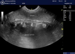
longitudinal plane view from the left flank showing a dilated, fluid-filled gastric fundus
No part of the small intestine was abnormally dilated. Blood flow within the cranial mesenteric vessels was demonstrated:
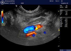
longitudinal plane view of the jejunal lymph nodes and intervening mesenteric vessels with colour Doppler
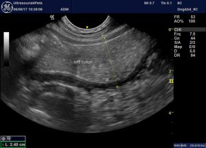
However, the colon was full with liquid content and thickened, hypoechoic wall:
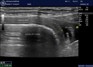
Furthermore, a small effusion was concentrated around this section of gut:
Following this bowel caudally the lumen tapered abruptly to a point. Here the gut wall could be followed around a tight circle exiting into an empty rectum:
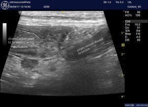
long axis view of the descending colon with a dilated ‘upstream’ part and an empty ‘downstream’ section to the right of image.
Detail of the empty rectum:
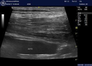
The twist is best appreciated in video
The site of torsion coincides with the root of the mesocolon and the caudal mesenteric vessels within the mesocolon are thrombosed:
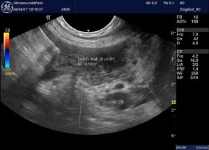
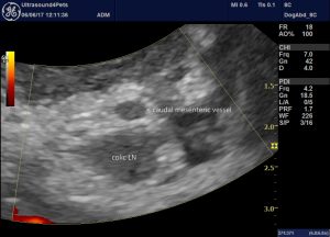
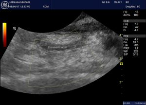
On the basis of a likely colonic torsion she was taken to laparotomy. A torsion of the entire remaining colon was confirmed; colectomy and ileo-colic anastomosis were performed.
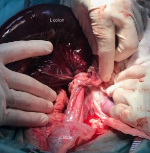
Recovery was relatively uneventful apart from a period of frequent VPCs managed with intravenous lidocaine.
Several points are noteworthy:
- German Shepherds have a well-documented predisposition to catastrophic visceral displacements.
- A history of prior gastrointestinal disease is quite common.
- Systemic sequelae and survival rates for colonic torsions in dogs are, perhaps surprisingly, often less severe than for mesenteric volvulus.
- two forms of colonic volvulus are described: where the caecum and ICCJ are involved the cranial mesenteric vessels are often also compromised. However, torsion of the tranverse and descending colon, as here, may leave cranial mesenteric flow unaffected.
- Although, as far as I’m aware, there are no detailed descriptions of colonic volvulus in the literature (all published cases appear to have been suspected from radiographic findings and confirmed at laparotomy) it does seem that findings in this dog were pathognomonic.


