Chronic intramural duodenal foreign body in a Dalmatian dog
Another slightly unusual case: a 3 year-old Dalmatian with a 5-month history of vomiting and weight loss.
On ultrasonography the stomach is dilated despite a 12-hour fast:
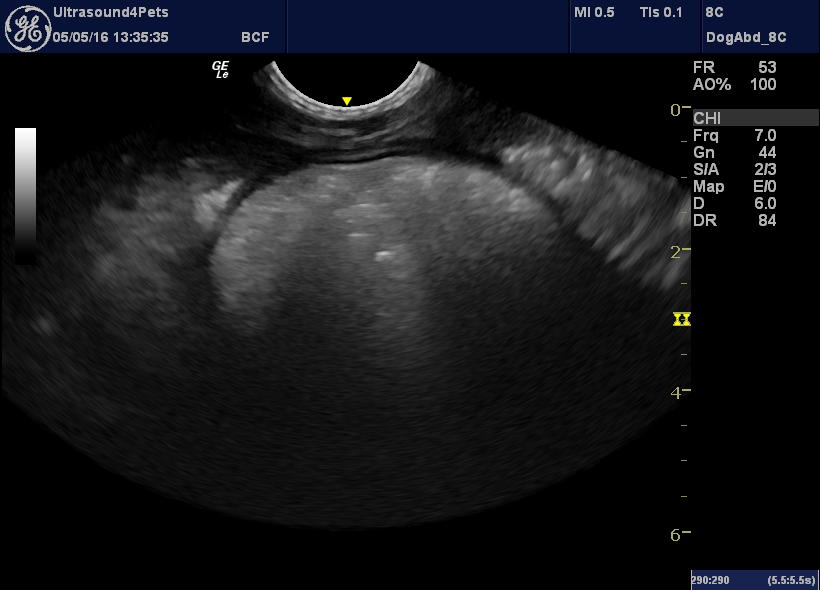
longitudinal pane view from the ventral mid-line of the body of the stomach
The area of the cranial duodenal flexure is also abnormal:
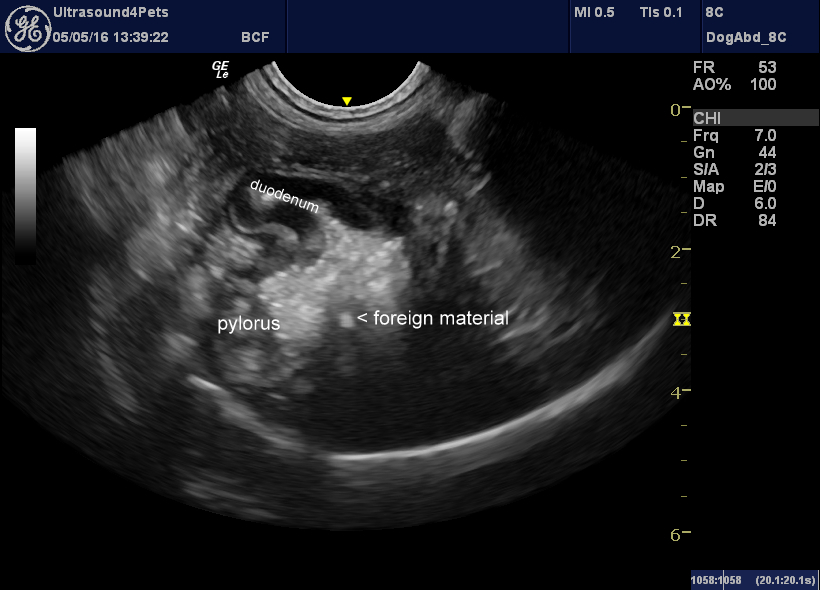
oblique view from the right paracostal area of the pylorus and duodnenal flexure
We couldn’t be sure at the time but there does appear to be a hyperechoic fragment of foreign material within the wall at this point.
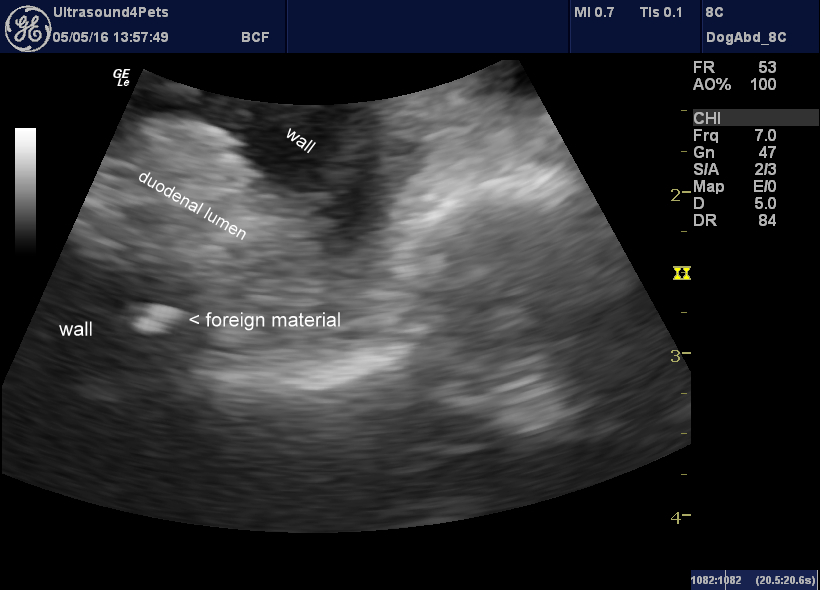
The adjacent duodenal lymph node is enlarged:
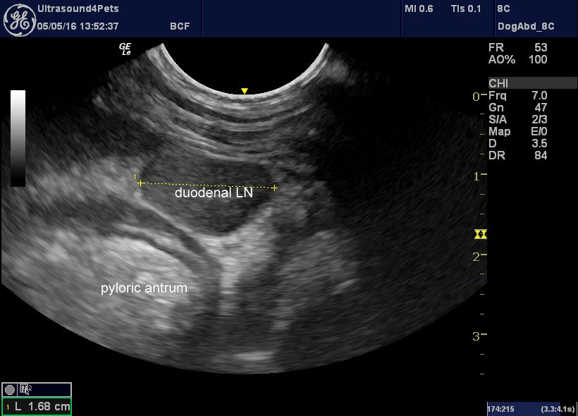
And there is dilation of the common bile duct implying obstruction at the level of the papilla:
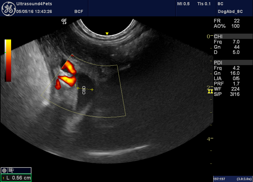
Given the chronicity and involvement of the duodenal papilla we weren’t optimistic. However, after some surgical heroics courtesy of Jon Mills at West Midlands Referrals the suspect section was excised and the pylorus/duodenal flexure reconfigured to ameliorate stricturing.
Histopathology revealed the presence of plant material and bacteria within the excised duodenal wall. At the time of writing the patient is recovering well.


