Chronic gastric volvulus in an old Weimaraner
This was a real challenge. A 13 year-old male Weimaraner with a history of gradual deterioration, intermittent vomiting and weight loss who then deteriorated over the course of a few days with more frequent vomiting or unproductive retching. On physical examination he was thin, alert but weak and with moderate abdominal pain. Femoral pulses were perhaps slightly weak and an intermittent tachydysrhythmia was noted. However, there was no overt abdominal bloating.
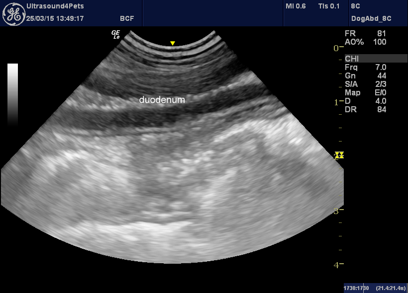
On ultrasonography most of the abdomen was really unexpectedly unremarkable. The first clue that something was amiss in the cranial abdomen was that a prominent loop of small intestine with mucosal thickness consistent with duodenum was present immediately deep to the abdominal wall on the left side.
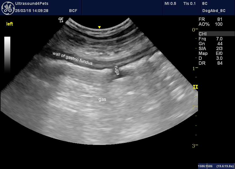
Initially it was difficult to get to grips with what was happening with the stomach. Clearly there was a lot of gassy content and the wide spacing of the rugae indicates distention:
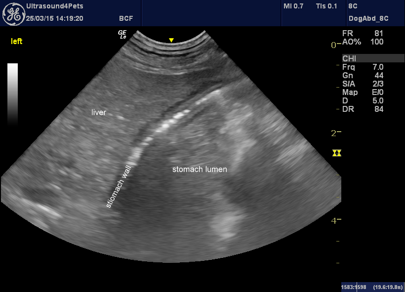
Exploring different viewing angles it was eventually possible to find a section of stomach wall adjacent to the liver with clearly abnormal wall -with intramural gas densities. This is a really unusual and concrete abnormal finding.
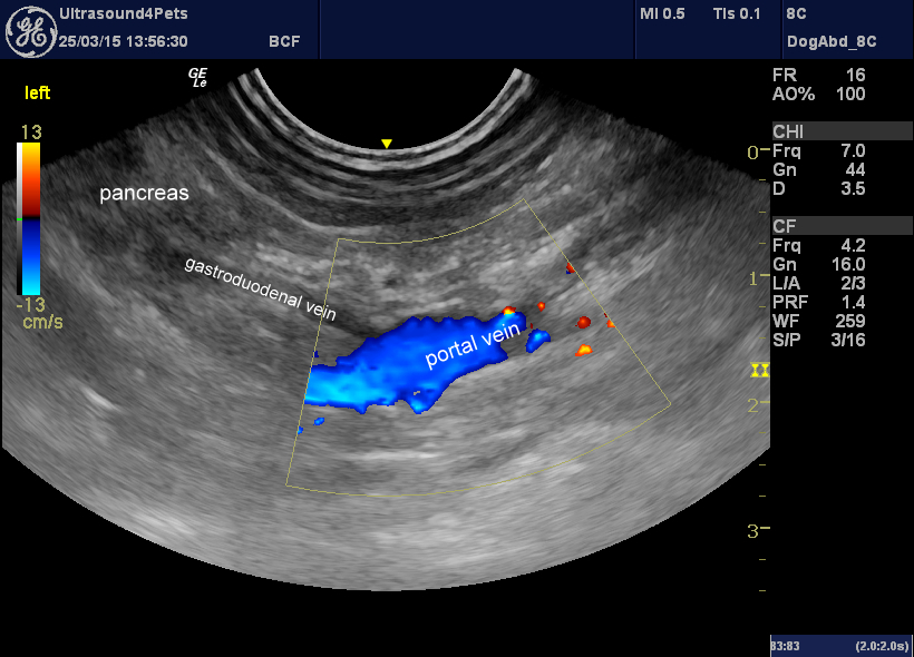
Signs are pointing to the possibility of gastric dilation-volvulus. However, this is a dog with no obvious external signs of bloat who has been poorly for several days. The portal vein remains patent (in full ‘classic’ GDV it is usually occluded):
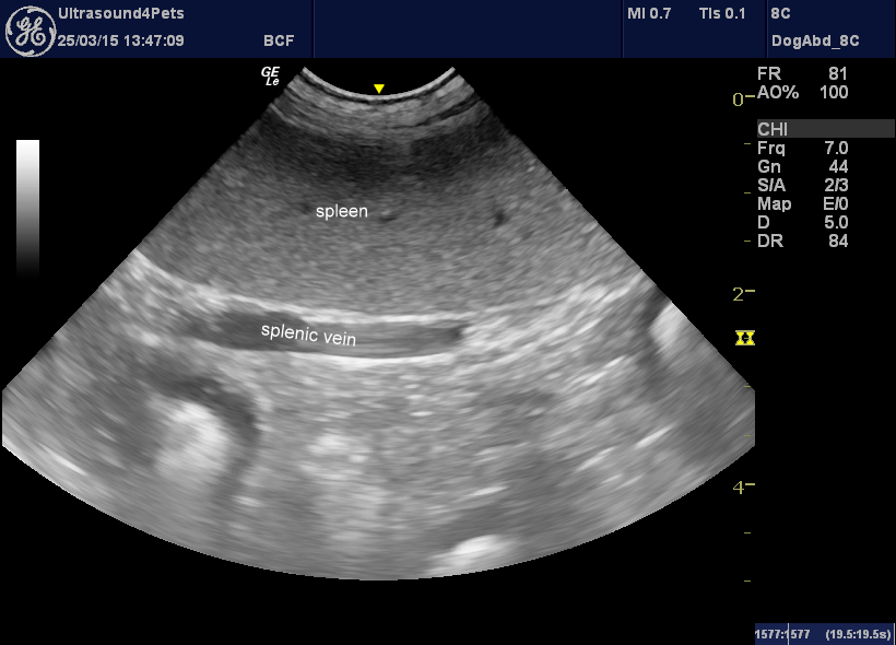
The splenic itself is unremarkable but the splenic veins are somewhat distended and spontaneous echocontrast is visible in the lumen:
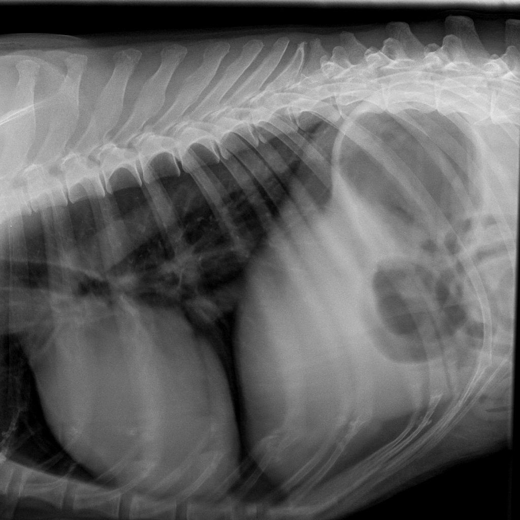
With a provsional diagnosis of chronic gastric volvulus we moved swiftly to xray:
There is clear compartmentalisation of the stomach with a ‘shelf’ consistent with volvulus. The degree of dilation is not extreme -this and the deep-chested conformation are presumably contributing to the lack of externally-obvious bloat.
Given his age and condition the decision was made for euthanasia.


