Canine patent ductus arteriosus (PDA) echocardiography
This is nothing very radical but when I started echo-ing I struggled for ages to get my brain around how to visualise PDAs. The degree of visibility does depend a bit on the individual dog’s conformation and how closely the main pulmonary artery and aorta are in approximation. This particular dog: a Springer Spaniel gives especially nice views.
Left cranial long axis view (cranial to right of image):
With the probe in the left armpit aligned with the long axis of the heart, the image which usually presents itself is of the ascending aortic root:
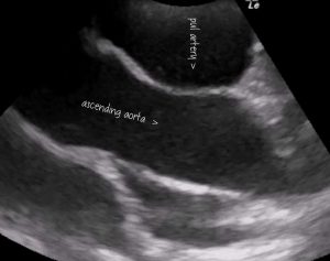
Freeze frame from the above video. The ascending aorta runs across the middle of the image. As seen in this orientation the LV is to the left and flow is directed from left screen to right through the aorta. The main pulmonary artery is wrapped around the aorta.
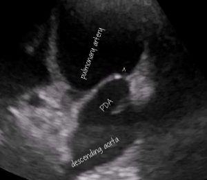
The video clip follows the aortic arch. As it bends around the front of the pulmonary artery the PDA comes into view carrying flow back up to enter the PA on its cranial aspect. To achieve this view rock/fan the probe to direct the beam more dorsally away from the ascending aorta whilst sliding the contact point towards the sternum. In some round-chested dogs the best view is with the probe almost on the sternum.
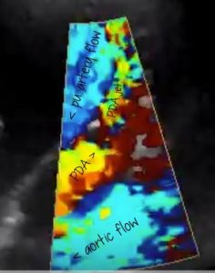
Same view as previous image with colour Doppler. Direction of flow in various parts according to arrows.


