Feline dermatology quiz
A little challenge for you all. What is this condition? He is a young DSH cat and both ears are affected. I’ll give it a few days. No prizes, just the glory!… Continue reading
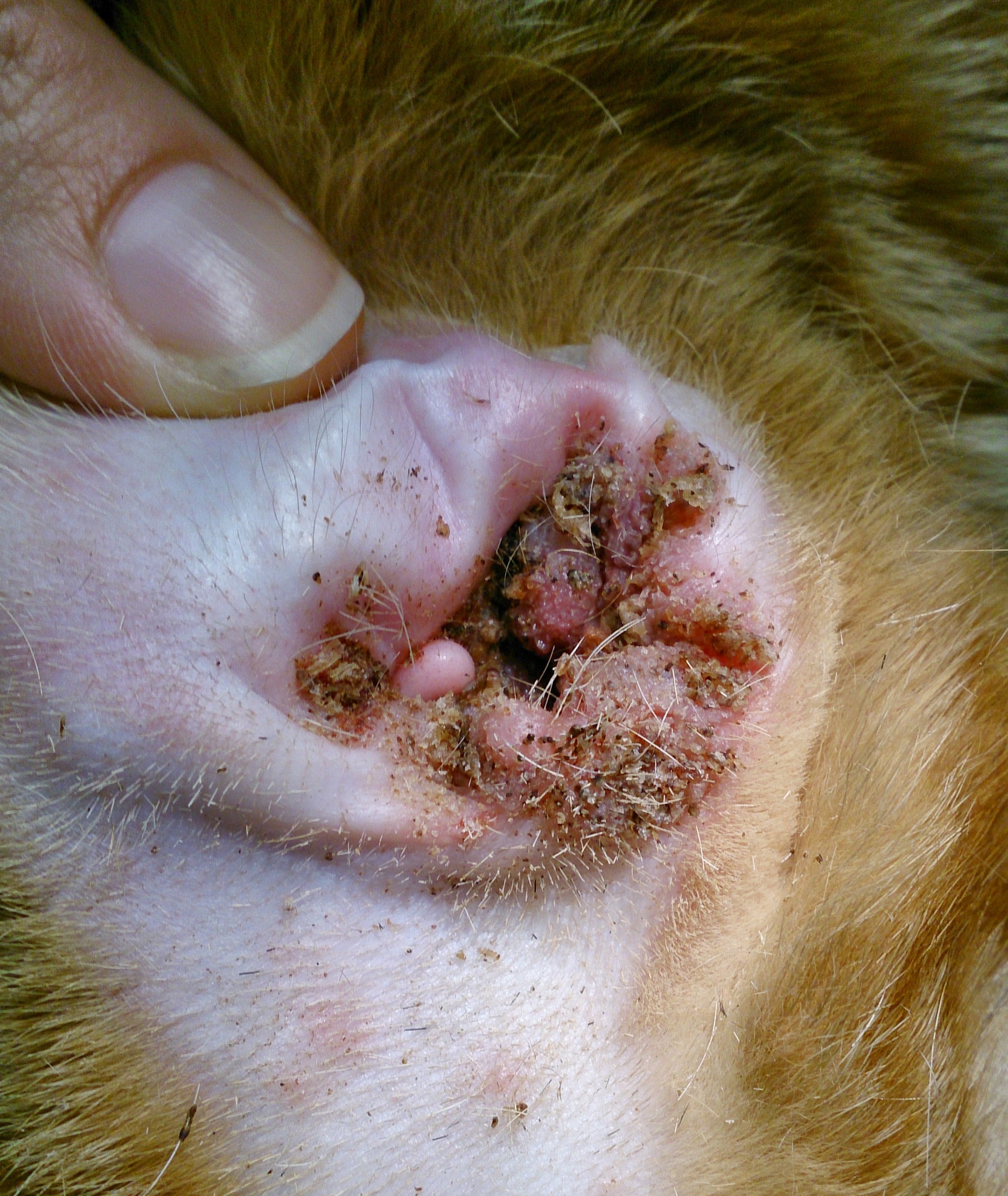
A little challenge for you all. What is this condition? He is a young DSH cat and both ears are affected. I’ll give it a few days. No prizes, just the glory!… Continue reading
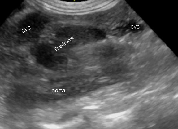
Adrenal ultrasonography is the guitar solo of canine and feline scanning: the bit that everybody wants to be able to do and have on their CV. Maybe adrenals and pancreas. And it’s true that if you can consistently find both adrenals then it gives you the confidence to book in those abdominal examinations. This little … Continue reading
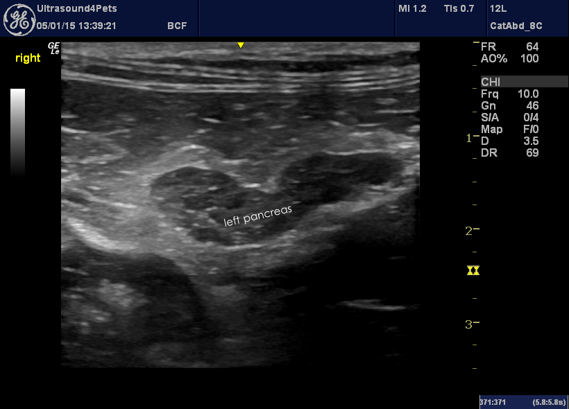
Just a few pictures from the last week to illustrate just how common pancreatitis is in both cats and dogs….and how much difference a high-frequency linear probe makes. This is the left pancreatic lobe from a cat with chronic recurrent abdominal pain. The pancreas is ringed by inflamed abdominal fat and the pancreatic parenchyma itself … Continue reading
This is just the most unbelievable story I’ve come across in some time. This 11 y.o. dog presented with episodic collapse. After a thorough work-up it transpired that she was sometimes hypoglycaemic and an insulin assay confirmed that levels were way above what would be expected at the time of hypoglycaemia. She was treated with … Continue reading
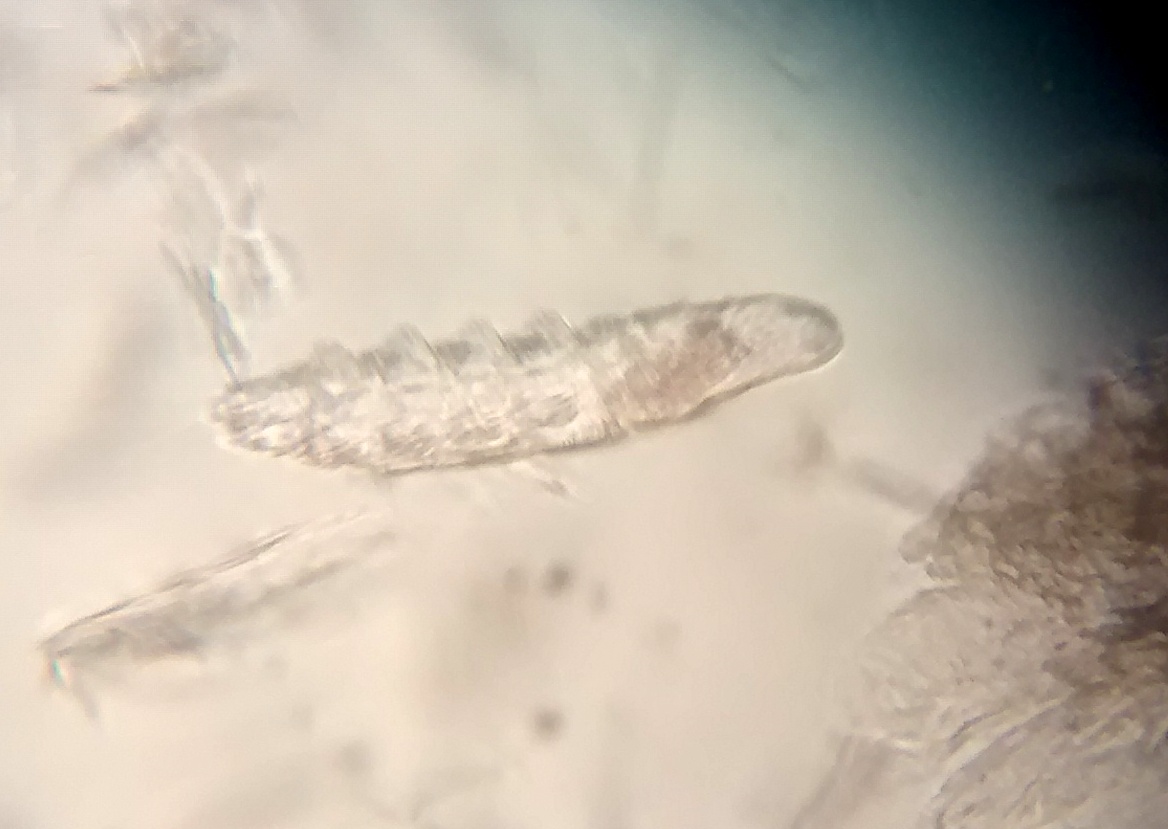
Continuing with demodicosis: most host species seem to carry two or three forms of this mite. In dogs, the long-bodied form D.injae inhabits the sebaceous glands and is associated with dorsal seborrhoea in terriers which is often pruritic. The ‘normal’ form D. canis spends most time lying in the hair follicles and the short-bodied (as … Continue reading
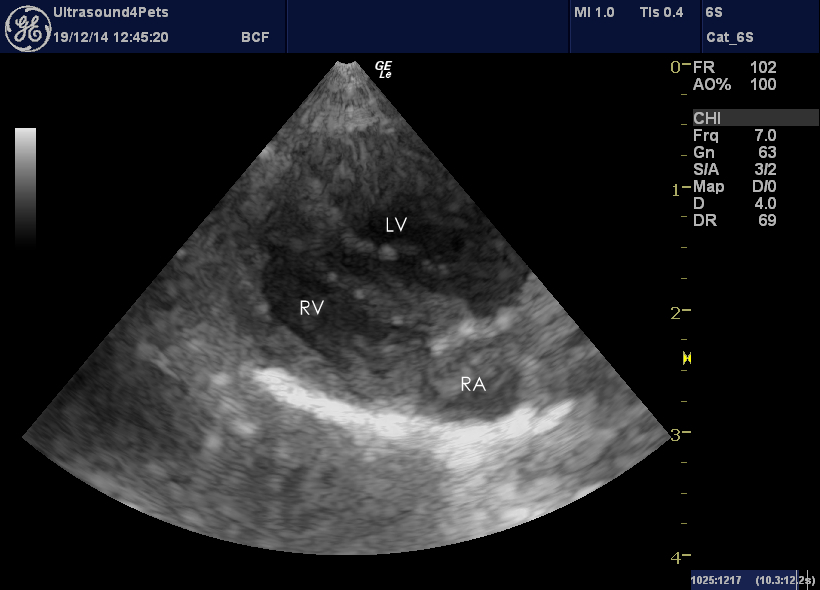
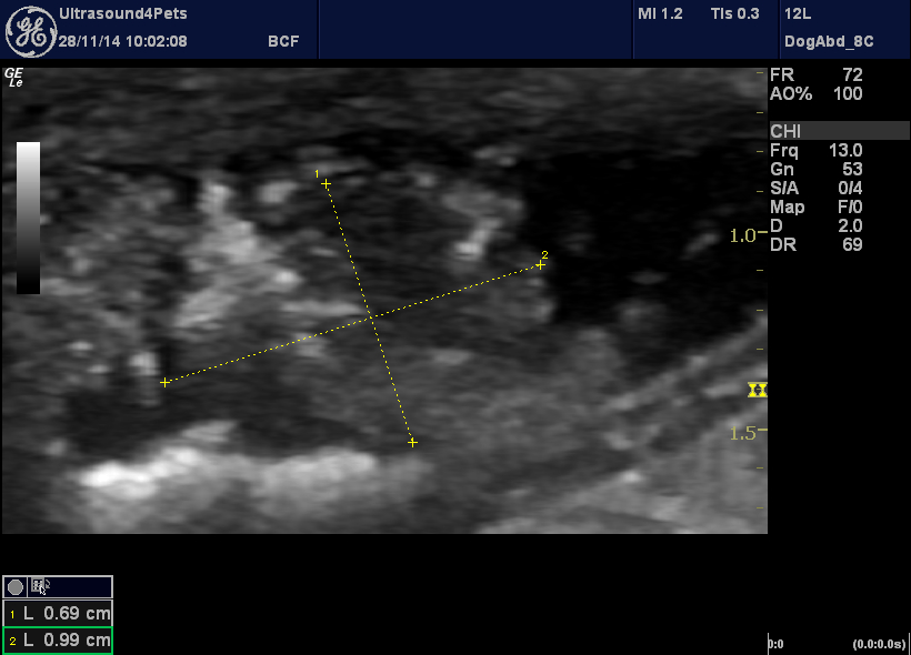
The thoracic medicine theme continues with this 3-month-old terrier pup. Initially presenting with stunted growth, matters then took a turn for the worse with sudden onset severe dyspnoea. On ultrasonography the heart was unusually difficult to see from the right side due to interposition of lung. From a left apical view the right ventricular wall … Continue reading
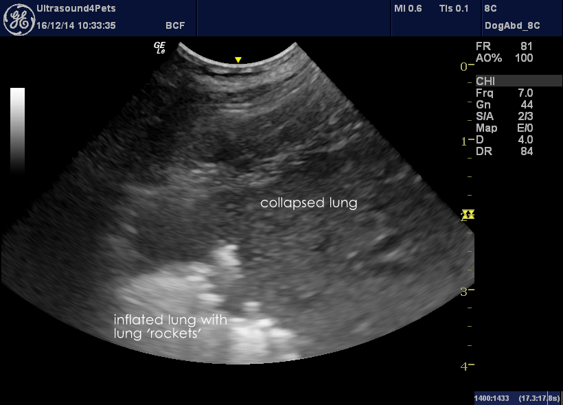
This is a really good recent paper on the subject from human medicine: Emerg Med J. 2012 Jan;29(1):19-23. doi: 10.1136/emj.2010.101584. Epub 2010 Oct 28. Lung ultrasound is an accurate diagnostic tool for the diagnosis of pneumonia in the emergency department. Cortellaro F1, Colombo S, Coen D, Duca PG. In summary these authors found that, in … Continue reading
We have just completed a prospective series of 48 canine acute abdomens examined ultrasonographically over the last 6 months. The main findings: 1: 8/48 were foreign bodies plus 1/48 entrapped hernia -more obstructions than I was expecting. That is maybe the most important single finding: that we almost certainly didn’t miss any. All of those … Continue reading
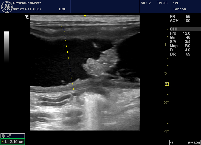
Now, this is a common problem. Dogs with dysuria and/or haematuria. You perform a full abdominal ultrasonogram and, behold! – an apparent soft tissue mass in the bladder. So, what is it? An FNA is generally regarded as being inadvisable since transitional cell carcinomas may seed along the needle line. Cystoscopy is certainly an option … Continue reading
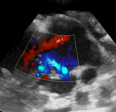
It always makes me mad when exam questions ask ‘What is the commonest…….’ because it varies so much from one publication to another and from one geographical area to another. Dysplasia of the tricuspid valve is supposedly relatively rare in cats; but it has been the most frequently seen feline congenital heart defect in our … Continue reading


