Apparent Sweet’s Syndrome in a dog: a steroid-responsive, sterile neutrophilic dermatitis
A two-year-old, neutered female Cocker Spaniel presented with a history of several days’ malaise, mild diarrhoea, inappetence and fever (40 C). Before any drugs were administered she then developed multiple widespread skin lesions consisting of sharply-demarcated ulcers up to 3cm diameter with swelling, overlying crust and surrounding erythema.

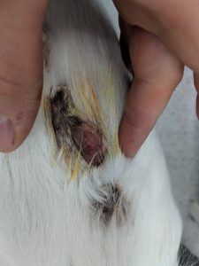
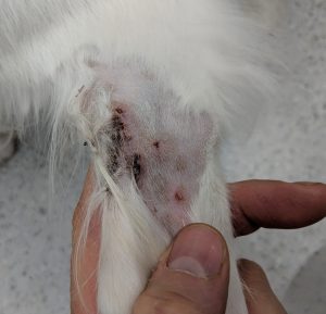
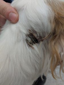
Figures: a) profile of the patient -note the degree of swelling of the raised dorsal lesion
b) ulcer on the dorsum
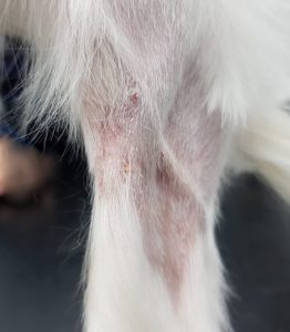
c) lesional skin of the hock: erythematous macules with central ulceration
d) dorsolateral neck: crusted ulcer
Mucous membranes were unaffected and physical examination was otherwise unremarkable. Full abdominal and thoracic ultrasonography also revealed no further pathology.
Blood biochemistry was unremarkable. Haematology revealed a mild non-regenerative anaemia (Hct 30%) and subsequent bone marrow biopsy findings were of erythroid hypoplasia and myeloid hyperplasia considered consistent with anaemia of inflammation/chronic disease.
Histopathology:
Findings are largely similar in all biopsies. They reveal the presence of a severe, predominantly neutrophilic infiltration of the dermis, with fewer histiocytes and scattered eosinophils, lymphocytes and plasma cells. There is prominent ulceration in some areas, and there is prominent oedema. In some of the biopsies, the infiltration is predominantly perifollicular, and multifocal neutrophils and occasional eosinophils are seen in small vessels. The ulcerated epithelium is covered by a thick crust composed of cellular debris, haemorrhage and degenerate neutrophils. In a few areas, there is crust formation with orthokeratotic hyperkeratosis, multifocal degenerate and non-degenerate neutrophils and scattered acantholytic keratinocytes. In very few areas, there is transmigration of the vascular walls by neutrophils, and occasionally vasculitis is suspected. There is occasional microhaemorrhage in some areas.
This is obviously all a bit unusual. Most of the acute ulcerating skin diseases of dogs (severe deep pyoderma, vasculitis, immune-mediated diseases and necrolytic disorders) can be ruled out, or are at least highly unlikely, on the basis of histopathological findings.
Clinical findings (specifically fever, systemic illness) and histopath would support a diagnosis of canine Sweet’s syndrome:
Br J Dermatol. 1964 Aug-Sep;76:349-56.
AN ACUTE FEBRILE NEUTROPHILIC DERMATOSIS.
SWEET RD.
https://www.ncbi.nlm.nih.gov/
There are fewer than ten reported cases of this condition.
Can Vet J. 2010 Dec; 51(12): 1397–1399.
Canine sterile neutrophilic dermatitis (resembling Sweet’s syndrome) in a Dachshund
Malcolm J. Gains, Andréanne Morency, Frédéric Sauvé, Marie-Claude Blais, and Yannick Bongrand
https://www.ncbi.nlm.nih.gov/
Vet Dermatol. 2009 Jun;20(3):200-5. doi: 10.1111/j.1365-3164.2009.
Extracutaneous neutrophilic inflammation in a dog with lesions resembling Sweet’s Syndrome.
Johnson CS1, May ER, Myers RK, Hostetter JM.
https://www.ncbi.nlm.nih.gov/
In several of these there was a possibility of adverse drug reaction as a causative or complicating factor.
In man, criteria for a diagnosis of Sweet’s syndrome include:
- Acute onset erythematous/violaceous plaques or nodules
- neutrophilic dermatitis
- preceding fever
- good response to corticosteroids
Although the cause remains poorly understood, many human cases seem to run a course involving an initial respiratory or gastrointestinal infection (as in this case) followed by a presumed immune-mediated reaction which can be ameliorated by treatment.
Having failed to improve with co-amoxiclav antibiotic treatment in the interim, the present case responded very promptly to prednisolone at 1mg/Kg sid po:
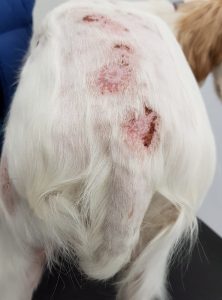
After four days of prednisolone the ulcers are dramatically improved


The pathologist who read my dog’s skin biopsy believes that my dog may have Sweet syndrome.