Another colic-caval portosystemic shunt in a cat
This is a cat who was investigated early in life on account of episodic malaise and raised bile acids. At the time a provisional diagnosis of primary hypoplasia of the portal vein was made.
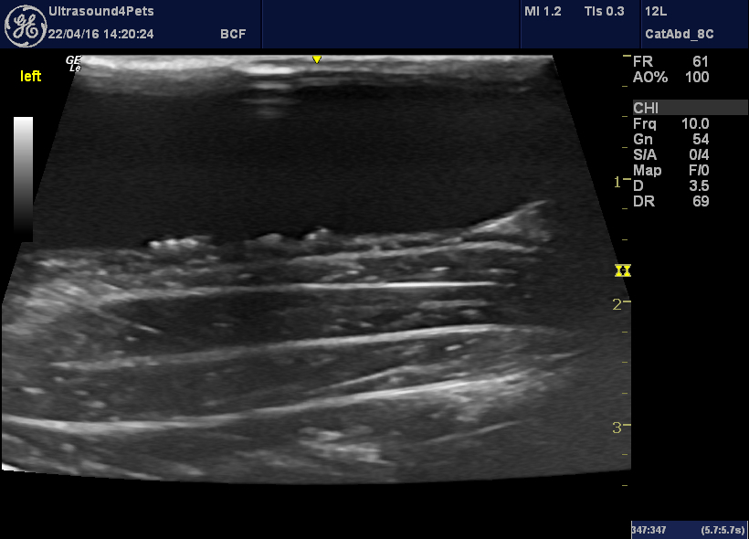
Small calculi in the bladder – a common finding in cats with portosystemic vascular anomalies (urates).
This is the main portal vein:
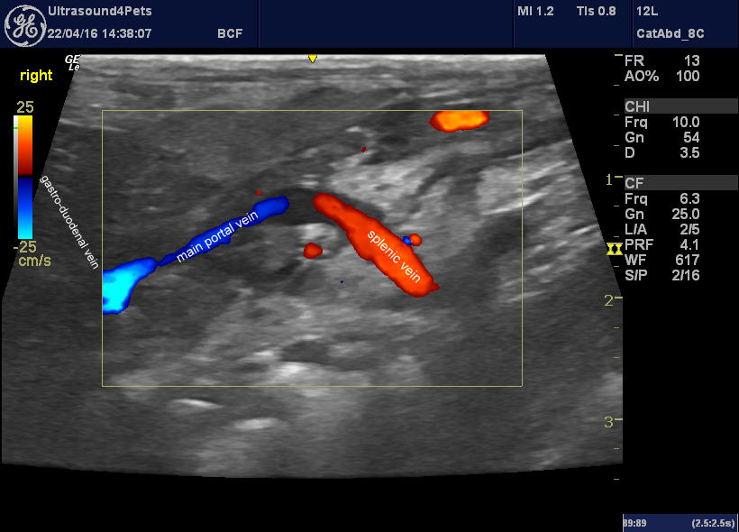
longitudinal plane view of the portal vein from the right side
The main portal vein is obviously and abnormally narrow but flow within it is hepatopetal. The splenic vein and gastrododenal veins also carry flow towards the liver. However, it’s not easy to find the mesenteric vein which should be joining the splenic vein to form the portal vein
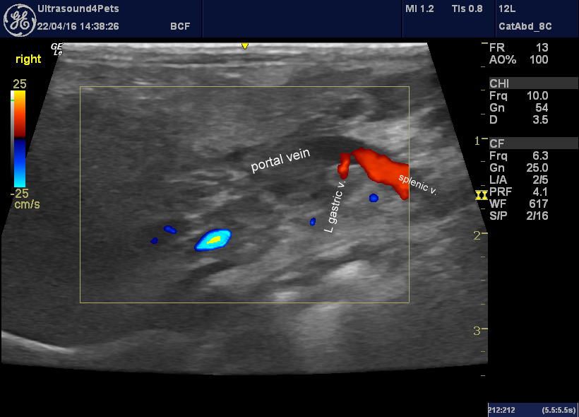
Further detail of the splenic vein as it enters the portal vein. The left gastric vein joins at about the same point.
.After a bit of a search, a markedly hypoplastic mesenteric vein can be found:
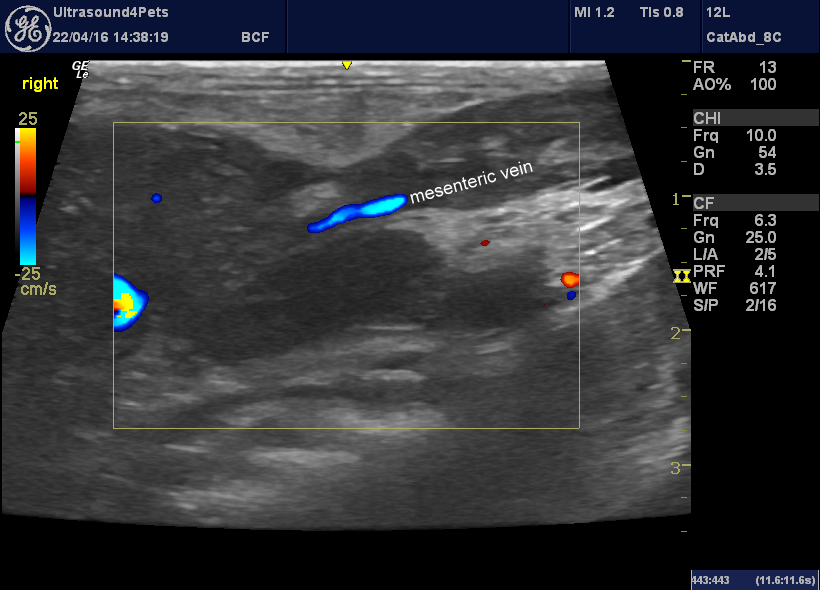
longitudinal plane view from the right side running towards the portal vein.
So, no obvious extrahepatic shunt in the usual sites (spleno-caval or porto-caval in cats) . But the suspicion is that most venous return from the guts is taking another route……
And this is the main contender -a large vein carrying caudally-directed flow which loops towards the pelvic inlet before turning 180 degrees and running alongside the cava and aorta:
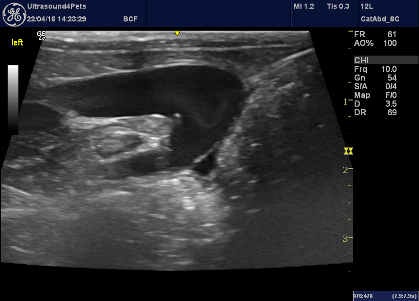
Longitudinal plane view from the left flank in the sublumbar area
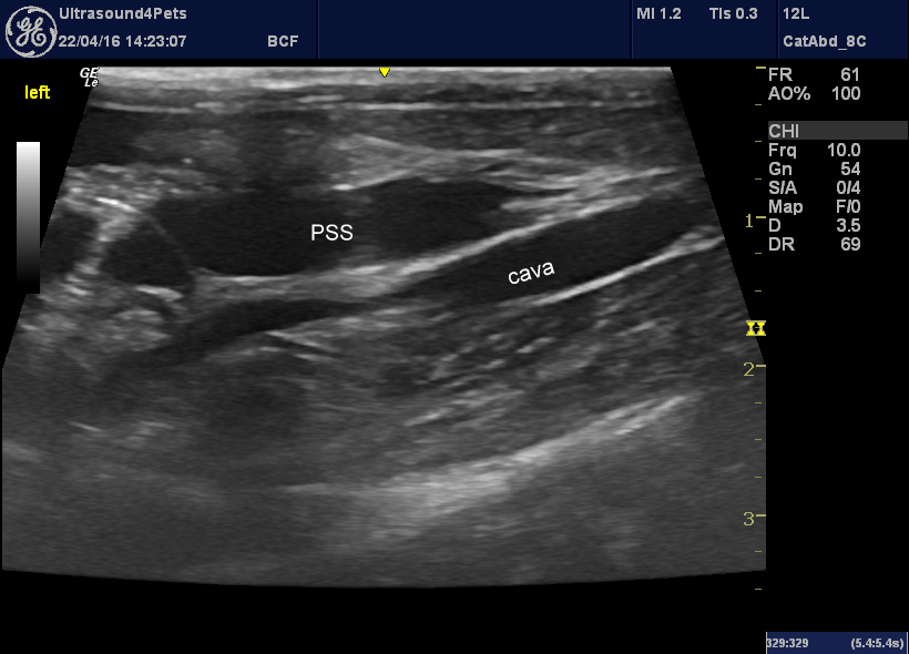
The presumed shunt running alongside the cava
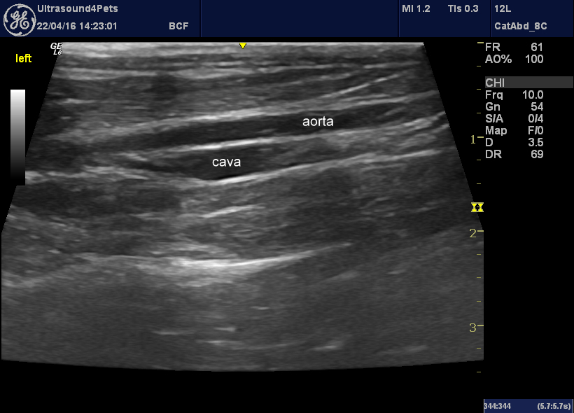
And just to prove that there are normal cava and aorta too
Finally it dives into the cava at the level of the left renal vein:
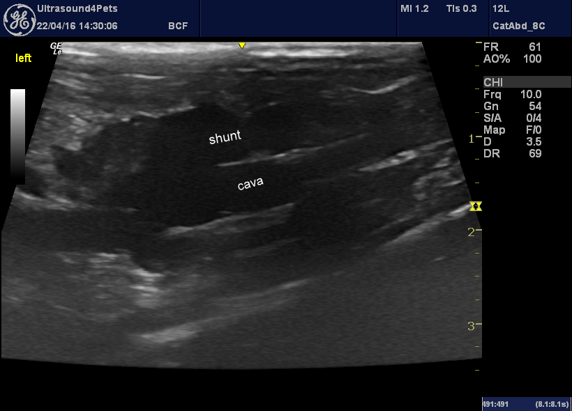
This appearance would be consistent with a congenital portosystemic shunt through the left colic vein (see images from the other case a few weeks ago).
Sadly this cat also has a nasty-looking pancreatitis: the main reason for current presentation.
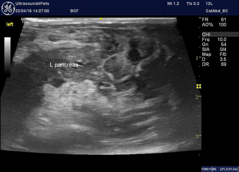
Longitudinal view from the left flank showing the mottled left pancreatic lobe with severe peri-pancreatic fat inflammation.


