Anaphylaxis, sepsis and pancreatitis
We are liking this very current article:.
Comparison of clinical findings between dogs with suspected anaphylaxis and dogs with confirmed sepsis.
Walters, Andrea M.; O’Brien, Mauria A.; Selmic, Laura E.; McMichael, Maureen A. Journal of the American Veterinary Medical Association.9/15/2017, Vol. 251 Issue 6, p681-688.
This is an interesting issue… I strongly suspect that I have missed diagnoses of anaphylaxis in the past and I’d like to improve in this area because management choices make a big difference these acute cases. Broadly speaking, the most striking finding of Walters et. al. is the variety of problems manifesting in anaphylactic patients. These aren’t just dogs with acute hypotension.
As the authors of the paper report, many physical and laboratory findings are common to anaphylaxis and sepsis (and probably to pancreatitis too): essentially these are largely features of SIRS.
Hypotension is also common in all groups.
Thrombocytopaenia, petechiae/ecchymoses, GI bleeding and prolonged coagulation times (both PT and APTT) are common: essentially DIC-like phenomena.
Pleural and/or peritoneal effusions may be seen in both scenarios.
Liver enzymes are frequently raised; with a tendency for ALT to be higher in anaphylaxis (range 200-5800 vs 9-1600) and AlkP higher in sepsis(range 20-1700 vs 31-133).
Potentially useful distinguishing features are:
- acute collapse was much commoner in anaphylaxis
- rectal temperatures tended to be higher in sepsis (although they ranged widely, no anaphylactic case was > 38.8)
- abdominal pain was commoner in septic dogs: presumably because the source of sepsis was often intra-abdominal.
- liver enzymes; see above
Obviously it’s also important to look for foci of potential sepsis such as GI perforations, abscesses and the like. Any effusions should be subjected to cytology looking for bacteria.
However, if you throw pancreatitis into the mix then things are becoming quite complex.
A recent case see by us was a 7 y.o. Yorkie presented with acute collapse, vomiting, abdominal pain, tachypnoea and tachycardia. Rectal temperature was 38.3.
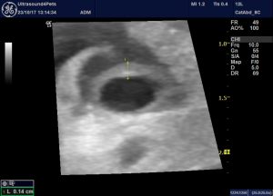
Systolic blood pressure was found to be 5o mmHg. The extent of hypovolemia can be seen on echocardiography:
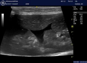
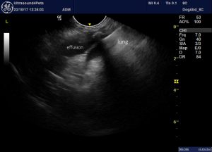
She exhibited bi-cavitary effusions:
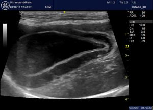
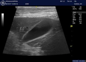
and dramatic oedema of the gallbladder wall:
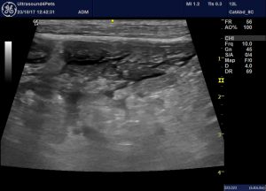
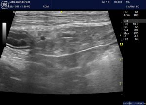
She also has pancreatic changes:
This is a very problematic area: I’m no longer confident that I can reliably distinguish ultrasonographically between pancreatic oedema and pancreatitis. The more hypoechoic the parenchyma and the more hyperechoic the surrounding fat then the more I’m inclined to pancreatitis but there is overlap. An abdominal effusion will always tend to make abdominal fat look hyperechoic.
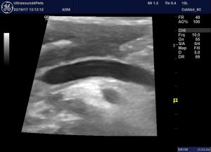
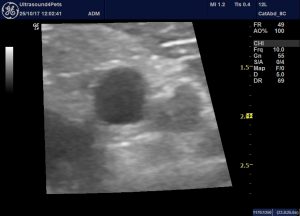
Her portal vein wall is distinctly thickened:
I suspect that this is a relatively specific sign of anaphylaxis: it makes a lot of sense that the inflammatory mediators are flooding into the PV from their main source in canine anaphylaxis -the gut.
Anyway, there were no visible bacterial in her abdominal effusion. She responded fully to fluids and dexamethasone (without antibiotics) and 48 hours later her corresponding images looked like this:
Dramatic huh!