Advanced adrenal scanning manoeuvres
Adrenal ultrasonography is the guitar solo of canine and feline scanning: the bit that everybody wants to be able to do and have on their CV. Maybe adrenals and pancreas. And it’s true that if you can consistently find both adrenals then it gives you the confidence to book in those abdominal examinations.
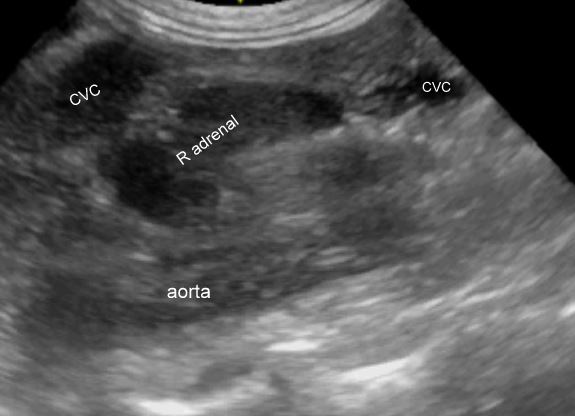
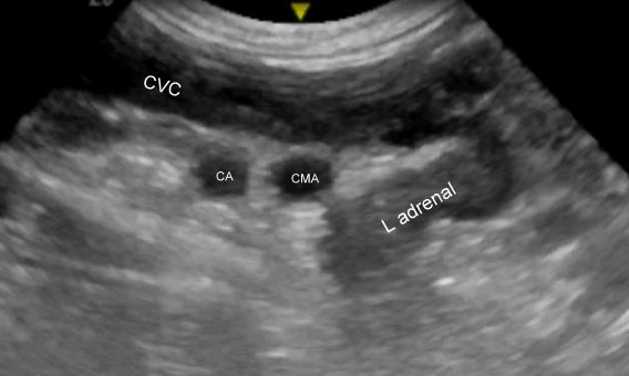
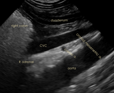
This little video clip is for ultrasound geeks and is intended to illustrate some of the useful landmarks. This clip was taken with the probe on the right side of the abdominal wall just under the last rib and in a longitudinal plane (left of screen is cranial). Both left and right adrenals are visible. Yes!… you can see them both from the same side. It’s not all that difficult to mistakenly assume that the adrenal-like thing which you are seeing is the one corresponding to the side on which you have the probe.
[wpvideo a4JBv4mr]
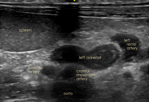
Crossing the top of the picture is the caudal vena cava (CVC). Below it is the aorta. Between them you can see the twin major arteries of the cranial abdomen -the coeliac (CA) and the cranial mesenteric (CMA). These structures are the key to locating the adrenals. The right adrenal lies immediately dorso-lateral to the CVC and just cranial to the coeliac artery. The cranial pole of the left adrenal is found just behind the cranial mesenteric artery.
In a crowded abdomen with difficult gas shadows you can get some orientation by applying a colour box to the area of the major vessels medial to the right kidney. The aorta, with its pulsatile flow usually sticks out like a sore thumb in the dorsal abdomen. Rotate the probe to optimise the aorta running across the screen and as you sweep along it you will see the paired CA and CMA exiting. The adrenals will be within a couple of centimeters of their origin.
A little colour (or power Doppler) also helps confirm that the structure which you identify as the likely adrenal really is a soft tissue structure and not a vessel. The hepatic artery branching off the coeliac runs cranially in the area dorsal to the CVC and can be mistaken for the right adrenal.
Part of the clinical significance of this right-sided view is that it reveals the relationship between the left adrenal and the CVC. In the normal view of the left adrenal from the left abdominal wall (below) the aorta, left renal artery and CMA are readily seen around the left adrenal but the CVC is not so obvious. This is important because left adrenal mass lesions certainly will invade the CVC if they can and it’s desirable to be able to demonstrate presence or absence of such behaviour.
A final hot tip…… From the right side, the aorta and CVC are easiest to,locate caudal to the kidney. There’s less gut gas in the way. As you follow them forward you reach the ‘dark zone’ created by the shadow of gas in the transverse colon and caecum.
Here, you can either try to massage the gas out of the way with your probe hand as you follow the vessels cranially; or you can just skip forward with mental extrapolation to where they emerge cranial to the shadow. The right adrenal sometimes lies in the shadow, sometimes just cranial and sometimes just caudal.


