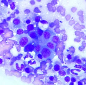
A further example of ‘Starry sky’ spleen
Following on from our splenic torsion case, a couple of days later I saw a similarly interesting spleen. The patient was a 9 y.o. MN mixed breed dog presenting with progressive malaise, anorexia, weight loss, abdominal pain and vomiting. On physical examination he exhibited ecchymoses/bruising on his flank.
His blood biochemistry was relatively unremarkable but he had a strongly positive Snap cPL test and moderate thrombocytopenia.
His abdominal sonographic findings are dramatic:
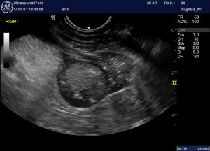
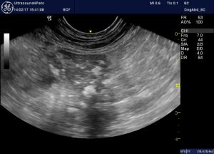
longitudinal view from left flank showing the spleen
There is an extensive part of the spleen with hypoechoic parenchyma interspersed with hyperechoic ‘lacy’ patterning. However, in contrast to the aforementioned splenic torsion patient the splenic change in this dog is confined to a discrete area:
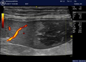
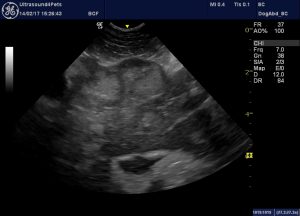
A similar view with power Doppler to demonstrate lack of perfusion in the abnormal area
And furthermore the hypoechoic area lacks blood flow on Power/colour Doppler. There is a rim of hyperechoic parenchyma at the margin of the avascular zone. There is no evidence that this is a mass lesion -the splenic silhouette is not disrupted. These findings would be consistent with an infarct.
The pancreas is grossly abnormal -being swollen and very heterogeneous. The peri-pancreatic fat (in fact the whole right cranial abdomen) is intensely hyperechoic consistent with inflammation.
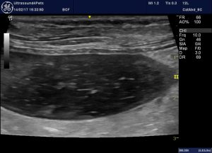
The tail of the the right pancreas as seen in longitudinal plane from the right flank
Sadly there is a scatter of ominous-looking mass lesions throughout the abdominal viscera:
Oblique view of right side of liver -there are multiple iscrete ‘target’ lesions -up to 3cm diameter