Ultrasound-guided Botox injection for canine cricopharyngeal achalasia
Hugo, is a Cocker Spaniel; about 5 years old. For months he had exhibited choking on swallowing and episodes of aspiration pneumonia. There was been no evidence of neurological dysfunction apart from dysphagia. Oesophageal appearance and function was unremarkable on radiography, ultrasonography and endoscopy. Ultrasonography of the upper oesophagus during feeding showed that very little food was making it through the upper oesophageal sphincter with each swallow; but each small bolus which did make it through was swiftly carried away caudally as normal.

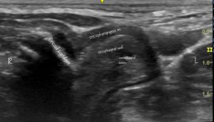
A Foley catheter was passed into the cervical oesophagus and the balloon inflated with sterile saline. When pulled cranially the balloon meets resistance at the level of the UOS and the cricopharyngeus muscle is seen encircling the oesophagus at this level.

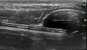
Longitudinal plane view of the UOS. with cranial to the left The inflated balloon and tube of the Foley catheter are easy to identify. With gentle traction on the Foley we were able to confirm that the stricture lay at the level of cricopharyngeus.
Using an out-of-plane technique (coming in from the side of the probe half way along) with a 5cm 22G needle we injected 10 units of Botox into the cricopharyngeus at various sites around ventral and lateral aspects of the UOS. I have tried finer needles in the past…but I find that they become hard to see.
Happily this seems to have been effective. After a few initial days of continued difficulty swallowing, signs have been much improved (up to the time of writing one month later).
Details of the technique from human medicine:
The technique has previously been reported in dogs; with mixed results:
https://www.koreascience.or.kr/article/JAKO201821142174136.pdf
Finally a little bit about in-plane and out-of-plane approaches to ultrasound-guided injection/vascular access which I like:


