Ultrasonography of pneumothorax
Ultrasonography is surprisingly good as a rapid, readily available imaging modality for screening potential pneumothorax patients. Since cardiogenic pulmonary oedema, pulmonary thromboemboli, pneumonia, pleural effusions and intrathoracic neoplasia can also be detected it’s a powerful tool.
The technique can be mastered by anyone with a few minutes to spare. Apply a micro-convex probe to the intercostal spaces and explore both hemithoraces systematically.
The normal appearance is of the chest wall in the near field bounded by a thin hyperechoic line marking the pleura. Obviously, the parietal pleura is normally in intimate contact with the lung pleura and the two slide against each other causing the ‘glide sign’.
In fact this dog’s lungs aren’t really completely normal because she has a large volume pneumothorax and those lungs are somewhat collapsed. This is causing the increased number of hyperechoic ‘B lines’ (see earlier posts) arising at the pleural interface.
These clips are recorded with the dog in a standing position. As the probe is moved dorsally a transition point is crossed where, on the inside of the chest wall, there is just free gas rather than lung. The glide sign disappears.
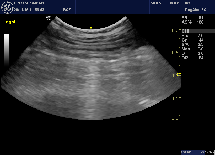
In still images the appearance is almost identical to normal lung.

longitudinal plane view through an intercostal space in a dog with pneumothorax showing the hypoechoic muscle of the chest wall in the near field and then the hyperechoic parietal pleura. The deep field consists of free gas. The appearance of still images such as this is almost identical to normal.
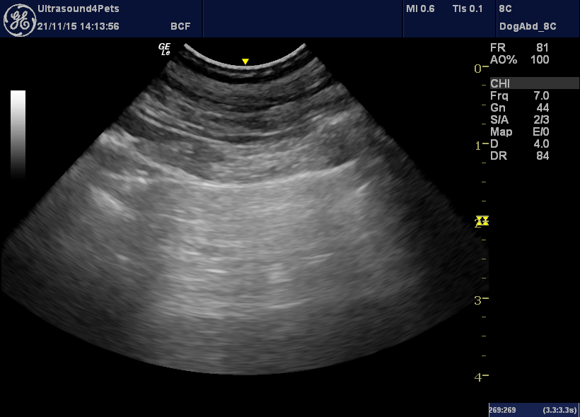
This is from another dog with properly normal lungs:

similar view from a dog with healthy lungs.
And a better video clip of the glide sign from a dog with healthy lungs -without the B lines.


