Lung ultrasound in the diagnosis of presumed pulmonary mycobacteriosis in a cat
Surprisingly, considering the wealth of information on conventional bacterial pneumonia, there is little published on the features of pulmonary TB in people.
Int J Environ Res Public Health. 2018 Oct; 15(10): 2235.
Potential Diagnostic Properties of Chest Ultrasound in Thoracic Tuberculosis—A Systematic Review
Francesco Di Gennaro,1,2,† Luigi Pisani,3,4,† Nicola Veronese,5 Damiano Pizzol,6,* Valeria Lippolis,4 Annalisa Saracino,1 Laura Monno,1 Michaëla A.M. Huson,7 Roberto Copetti,8 Giovanni Putoto,2 and Marcus J. Schultz3,7
https://www.ncbi.nlm.nih.gov/pmc/articles/PMC6210728/
…and even less in cats and dogs.
The present case is an 11 y.o. DSH with a history of chronic cough unresponsive to cefovecin or glucocorticoids. Routine culture of a bronchoalveolar lavage sample yielded no growth. Cytology of the same sample revealed a mixed inflammatory cell population but no specific agents were visible after modified Wright’s staining. Serology did not support a diagnosis of toxoplasmosis.
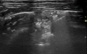
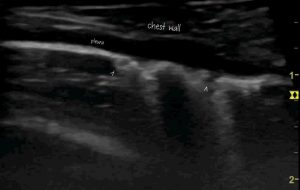
Lung ultrasound showed diffuse pleural thickening and a wide scatter of consolidated lesions in both hemithoraces. One larger lesion exhibited characteristics of the ‘shred’ sign normally associated with pneumonia -but in this case with heterogeneous echogenicity including some irregular areas.

More numerous, smaller lesions ranged from the lower limit of detectability at @1mm diameter and often coalesced into an irregular subpleural ‘plaque’.

Blood samples from this cat tested positive on interferon gamma release assay with specific response to antigens associated with less pathogenic strains such as M. microti.
This blood test, available through Biobest, may be worth considering in cats with chronic respiratory disease.


