Sonographic features of Leptospirosis in dogs in the UK
There’s a nice recent article on the subject of ultrasound findings in dogs with lepto:
Vet Radiol Ultrasound. 2018 Jan;59(1):98-106. doi: 10.1111/vru.12571. Epub 2017 Nov 1.
Prospective evaluation of abdominal ultrasonographic findings in 35 dogs with leptospirosis.
Sonet J1, Barthélemy A2, Goy-Thollot I2, Pouzot-Nevoret C2.
https://www.ncbi.nlm.nih.gov/pubmed/29094440
by
….but that was in France and, now, after Brexit we will obviously require UK sonographic criteria for the diagnosis of leptospirosis.
Renal changes were predominant in the French study: increased renal cortical echogenicity (100%), increased medullary echogenicity (86%), reduced corticomedullary definition (80%), cortical thickening (74%), renomegaly (60%), pelvic dilation (31%), and medullary band (14%).
This first patient of ours, a 7 year-old Jack Russell, suffered a very acute and severe, ultimately fatal, PCR +ve episode of leptospirosis. On presentation he was azotaemic (creatinine 400, BUN 23), ALT only 168, AlkP 5000, tbil 130, DGGR lipase 75 (ref <45).
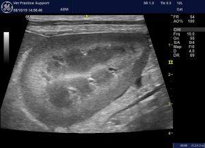
Left kidney of a Jack Russell Terrier with acute Leptospirosis 48 hrs after onset of signs
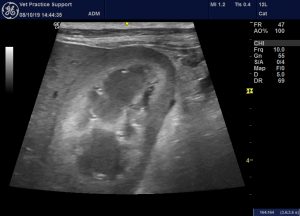
Same patient: right kidney
Now, I don’t know about you lot but I find renal echogenicity a lot more difficult to assess than is generally suggested by the regular texts. A 12 y.o dog’s kidneys will almost always look hyperechoic compared to a 1 y.o.. And a lot of ‘normal’ individuals will have some degree of subclinical nephropathy reflected in the appearance of their kidneys. It’s also true that sonographic architecture of the kidneys varies a lot with a few degrees of change of angle -especially in longitudinal plane views.
I’m not sure I would necessarily have judged those kidneys to have ‘abnormal’ echogenicity.
However, there is certainly a very marked anechoic, peri-renal and retroperitoneal effusion: which is obviously a very unusual finding in a dog.
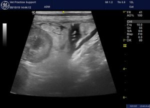
Retroperitoneal free fluid behind the right kidney.
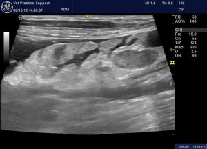
…and on the left side too.
They don’t all look like that. This is another PCR +ve case about one week after initial presentation:
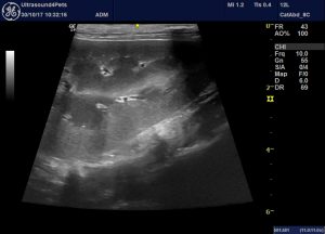
Apart from being towards the hyperechoic end of the spectrum of kidney echogenciity, I’m not sure this looks very abnormal
Next most frequently affected organ system in the French study was the liver: diffuse hypoechoic parenchyma (71%), hepatomegaly (60%). Biliary gallbladder abnormalities were found in 60% of the dogs, with biliary sludge (46%), wall thickening (29%), mucocele (26%), and hyperechoic wall (20%).
We would concur that diffuse hypoechoic liver parenchyma is typical:
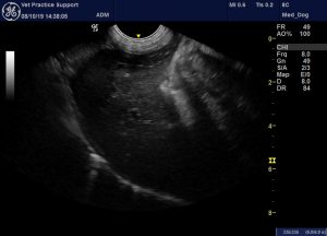
longitudinal plane view of the liver in a dog with acute leptospirosis: the hypoechoic appearance of the parenchyma in enhanced by contrast with diffusely hyperechoic abdominal fat.
On closer inspection, liver change is often heterogeneous:
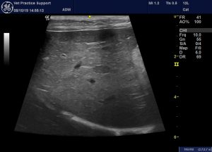
This is the same dog as in the previous image but with a linear probe. This shows how settings and probe dependent the appearance can be.
The gallbladder is often abnormal. I’ve reported this case before which I believe may have a blood clot in his gallbladder.
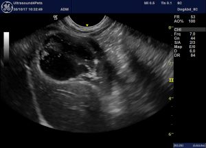
A putative haemocholecyst in an IFA +ve lepto case. The gallbladder lumen contains what is probably a blood clot.
Various gastrointestinal changes were reported by Sonet et al., affecting a high proportion of cases to some degree:
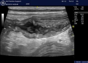
Long axis view of the left colon in leptospirosis: there is diffuse wall thickening with hyperechoic mucosa and liquid diarrhoea in the lumen
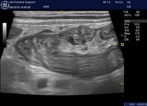
The ileo-caeco-colic junction: ileum (lower right of image) and colon are diffusely thickened
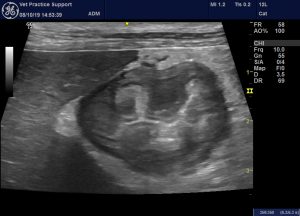
Longitudinal plane view of the stomach body. Again there is diffuse wall thickening and the abdominal fat is diffusely hyperechoic.
In a dog with these gastrointestinal changes and lacking dramatic renal or hepatic features it would be easy to overlook the possibility of lepto and call it another non-specific gastro-entero-colitis.
It’s interesting to note that the Leptospira serovars identified in the French dogs included a high proportion (e.g. serogroups Panama and Autumnalis) which do not match serovars included in either L2 or L4 vaccines in the UK. My understanding is that there is very little published information of the incidence or typing of clinical lepto cases in the UK.
The breeds of dog most affected match almost exactly our experience n northern England: German Shepherds, Jack Russell Terriers and Labradors in particular.
As another aside, in a recent literature review I could find no published evidence of beneficial effect of any antibiotic on disease progression in leptospirosis in any species (including people). This was acknowledged to be the case by several authors of meta-analyses. But you’d be brave not to…..!


