Lung ultrasound: ‘flooding’ in fulminant pulmonary oedema in cats and a comparison with pneumonia
Pulmonary oedema in cats and dogs is typically characterised by increased ‘B lines’ (also known as ultrasound lung rockets or comet-tail artefacts). This is a feature of oedema in the broadest sense and is not specific to aetiology. Differential diagnosis of this phenomenon includes trauma, electrocution, acute respiratory distress syndrome/acute lung injury, overhydration, asthma, near-downing and post-strangulation. However, left-sided congestive heart failure is the commonest cause of diffusely increased B lines.
Some care has to be exercised: normal cats often have a few B lines at the periphery of their lung lobes. The central parts are generally clear.

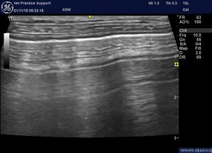
Nomal lung ultrasound image from a healthy cat: the brightest, most echogenic horizontal line is the pleurae. Above that in the image the lines represent fascial plane boundaries. Below it are horizontal ‘A lines’ which are artefacts -echoes of those superficial to the pleurae.
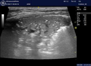
Cats, as ever, are not small dogs though. Really fulminant cardiogenic pulmonary oedema in cats can result in ‘flooding’ of one or more lobes.
That’s not something I’ve ever seen in a dog. Nor is it described in the excellent 2017 JVIM account of cardiogenic pulmonary oedema in dogs by Vezzosi et al..
J Vet Intern Med. 2017 May;31(3):700-704.
Assessment of Lung Ultrasound B-Lines in Dogs with Different Stages of Chronic Valvular Heart Disease.
Vezzosi T1, Mannucci T1, Pistoresi A1, Toma F1, Tognetti R1, Zini E2,3,4, Domenech O2, Auriemma E2, Citi S.
https://onlinelibrary.wiley.com/doi/full/10.1111/jvim.14692
This is a still image of the same area:

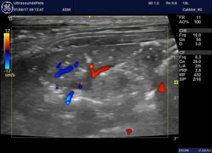
And with colour Doppler to show it’s not pulmonary thromboembolism as far as I can tell.

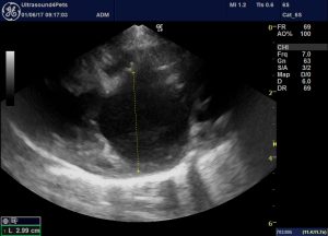
And the heart in this poor cat:

It’s worth comparing that video clip of flooded oedematous lung with this one from another cat with presumed pneumonia:
This was a young cat with acute onset tachypnoea, no murmur or gallop and unremarkable echocardiography who recovered within a week after treatment with antibiotics only and has suffered no recurrence of signs in the subsequent 18 months.
There is a small pleural effusion and the lung pleura bears a fine network of adherent presumed fibrin strands. The consolidated area is much more sharply-defined peripherally than in the CHF cat and exhibits the typical serrated boundary of pneumonic lesions. The consolidated area is more solidly hypoechoic than with oedema with fewer remnant air bubbles (although that varies from case to case). This cat also has large areas of sonographically-normal lung. Although local cardiogenic pulmonary oedema is occasionally encountered it’s not common to see a full spectrum from normal to completely collapsed/consolidated/flooded in the same cat.


Hi Roger, beautiful images. Really enjoying your website which I stumbled across a few months ago. I am curious re the flooding. It seems the lung is consolidated but I don’t see fluid as expected in the periphery (with B lines to infinity) w/severe oedema – there does appear to be fluid in the central part of the image/lung though. Bit confused as to how a flooded lung is diagnosed or classified. Do you have any links or references by any chance?
Many thanks
Raj Mukherjee MRCVS
Hi Raj, thanks for your interest! I wrote the post because there’s nothing i can find in the veterinary literature on cardiogenic pulmonary oedema progressing to the extent that the lung is completely flooded like this. I’ve never seen it in a dog -it seems to be a cat phenomenon. I think the B line phenomenon is due to reverberation within bubbles in oedematous lung isn’t it; but when the lung is completely flooded it just looks the same as consolidated lung. In human medicine the easiest comparison is with lung which has been deliberately flooded for diagnostic purposes e.g. https://www.ncbi.nlm.nih.gov/pubmed/29627084 Hopefully you can access full text of that via some route. The images show lung that looks just like pathologically flooded lung in the cat in the blog. no B lines. You can also access the images in that paper by googling ‘one-lung flooding ultrasound’ searching just for images. all the best, roger