Sonography of presumed granulomatous colitis in a French Bulldog
A severe colitis refractory to conventional treatments is not uncommon in young French Bulldogs and Boxers in the UK:
https://onlinelibrary.wiley.com/doi/pdf/10.1111/jvim.12020
A definitive diagnosis requires histopathology; conventionally using mucosal biopsies acquired at colonocopy. However, it’s certainly worth having a look with ultrasound before embarking on scoping. This helps to identify the worst-affected sections of gut.

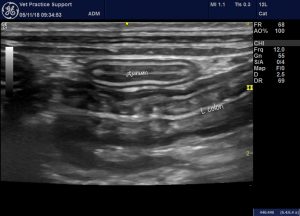
Long axis view of L colon: diffuse wall thickening

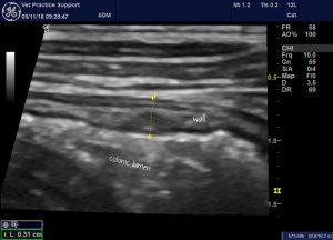
Detail of thickened colonic wall

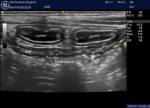
And a scatter of lesions in the submucosa of the descending colonic wall
These discrete, hypoechoic lesions in the colonic submucosa are clearly the same as described by Citi et al. in 2013:
Vet Radiol Ultrasound 2013;54(6):646-51
Micronodular ultrasound lesions in the colonic submucosa of 42 dogs and 14 cats
Simonetta Citi 1, Tommaso Chimenti, Veronica Marchetti, Francesca Millanta, Tommaso Mannucci
https://onlinelibrary.wiley.com/doi/abs/10.1111/vru.12077
…who identified these lesions in dogs and cats with a spectrum of gastrointestinal presentations and proposed that they represent lymphoid follicles.


Roger, those hypoechoic nodules in the sub mucosa of the colon are lymphoid nodules that are stimulated or reactive. Ulcerative colitis lesions extend into the mucosa. I have images if you want them, Bob.