Congenital lobar emphysema in a pup
The thoracic medicine theme continues with this 3-month-old terrier pup. Initially presenting with stunted growth, matters then took a turn for the worse with sudden onset severe dyspnoea. On ultrasonography the heart was unusually difficult to see from the right side due to interposition of lung. From a left apical view the right ventricular wall looks subjectively of similar thickness to the left. It’s not a great image -bear in mind that this is a 0.4Kg puppy!
Additionally, in transverse view, the interventricular septum is flattened to the left in late systole (the right ventricle is taking longer to empty than the left):
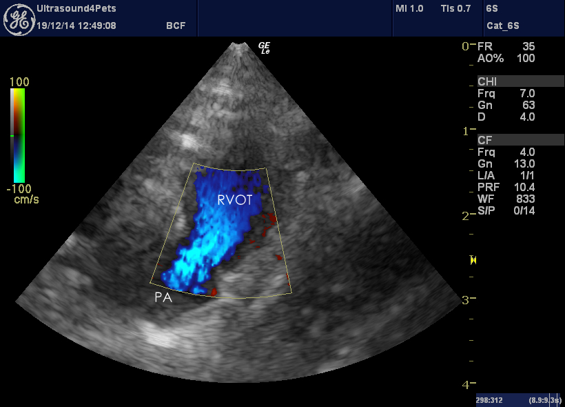
These both imply right ventricular pressure overload. And yet there is no evidence of any shunting or outflow obstruction: this transverse view of the right ventricular outflow tract (RVOT) and main pulmonary artery (PA) with colour shows nice uni-directional laminar flow.
With this being a tiny pup, gasping for breath we didn’t have a lot of time for assessing pressure gradients but we concluded that pulmonary hypertension was a likely cause. And so to xray….
And those lungs are not normal! The right diaphragmatic lobe is enlarged and hyperlucent. In contrast, the remaining lung lobes are somewhat collapsed and the heart is displaced cranially and to the left. Findings consistent with congenital lobar emphysema (CLE). Not entirely unreasonably, her owners opted for euthanasia rather than lung lobectomy.
CLE is an uncommon condition in which a congenital bronchial cartilage defect leads to ballooning of a single lobe in young dogs and resulting compression of the remaining thoracic contents. Terriers seem to be most often affected. There are only a few cases written up in the literature although we do see them from time to time so they may be under-reported. Although the condition is congenital the deterioration can be precipitous when lobar expansion really kicks in.


