Congenital Intrahepatic Portosystemic Shunts
These are a few recent canine examples. A prominent dilated venous structure in the centre of the liver is a frequent, relatively straightforward pointer to a likely diagnosis:
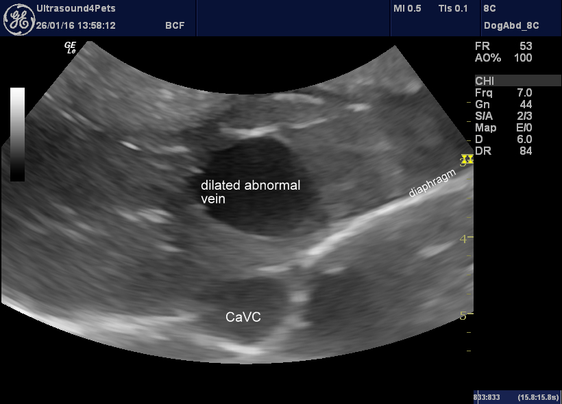
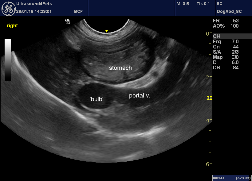
In this video clip the liver is seen in transverse plane. The hypoechoic structure nearer the top of the picture is the portal vein which flows into a dilated ‘bulb’ containing turbulent blood flow with spontaneous echocontrast. This communicates via a shunt vessel with the left hepatic vein and thence with the caudal vena cava (at the bottom of the image). The bright hyperechoic line across the image is the diaphragm.
This dog later underwent CT angiography (confirming it as being consistent with a patent ductus venosus) and surgical attenuation.
This is the same view as a still image:
And in longitudinal plane (the small liver size is more apparent in this view):
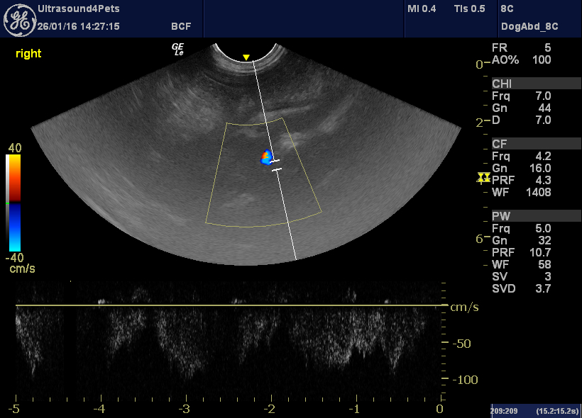
With PW Doppler, flow in the main portal vein is abnormally rapid -there is little resistance to flow as it zips straight through into the cava. And flow fluctuates with the respiratory cycle. This is a common feature where portosystemic communication (intra- or extra-hepatic) is present but not completely reliable -dogs which pant or breathe heavily during the examination may exhibit the same phenomenon.
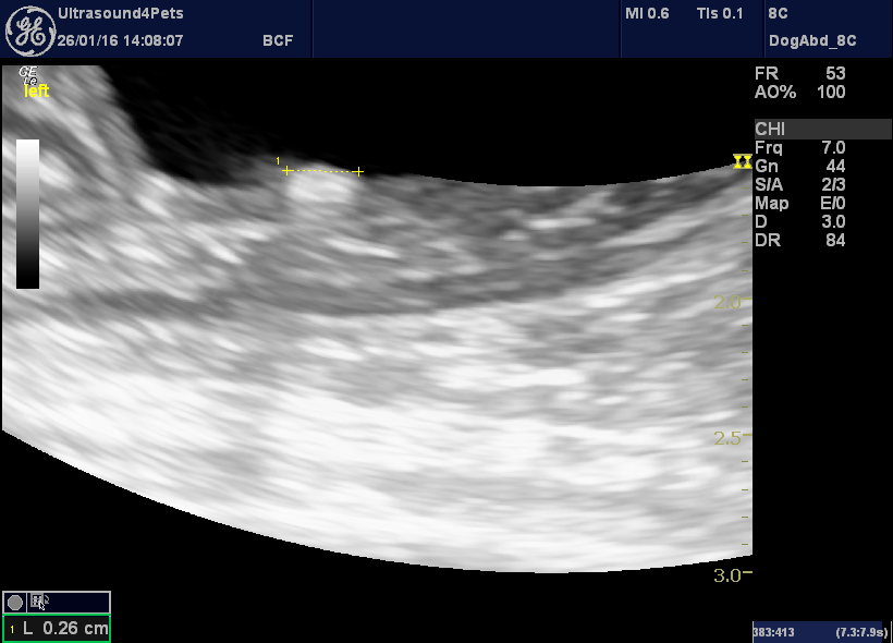
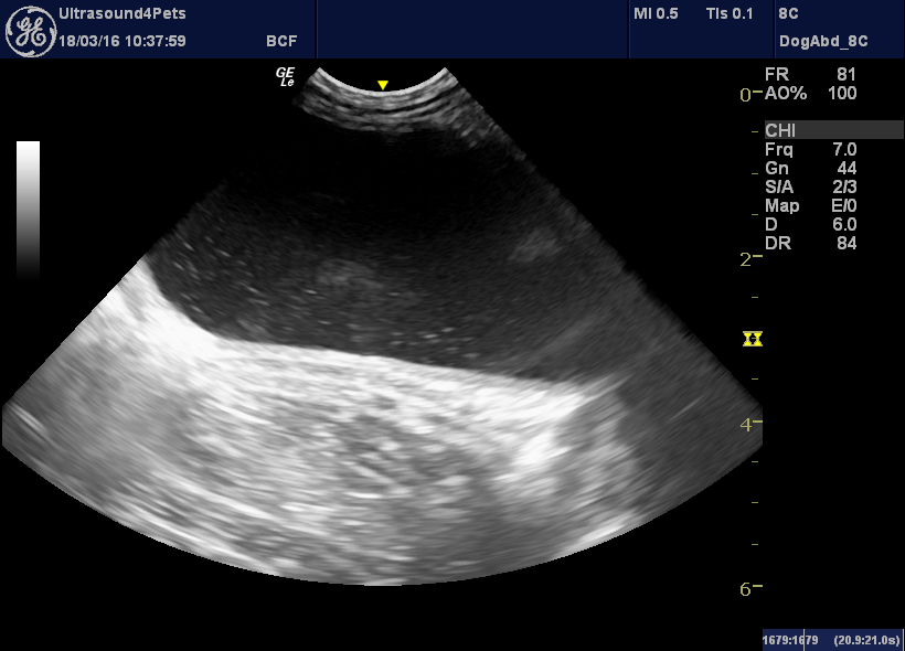
The presence of calculi in a young animal should always raise suspicions of there being a shunt (with resultant urate crystalluria).
This is a second, similar, dog ( a Border Collie):
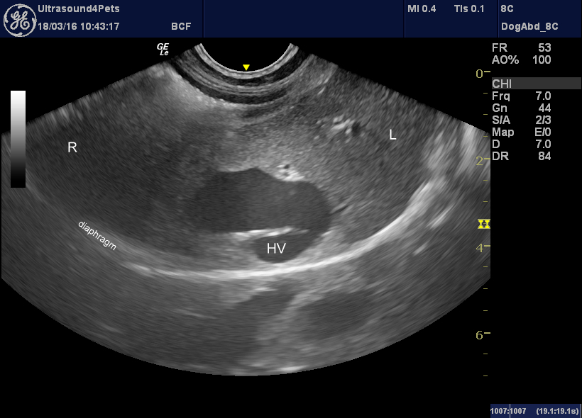
HV = left hepatic vein
Looks familiar doesn’t it?
….and portal vein flow….
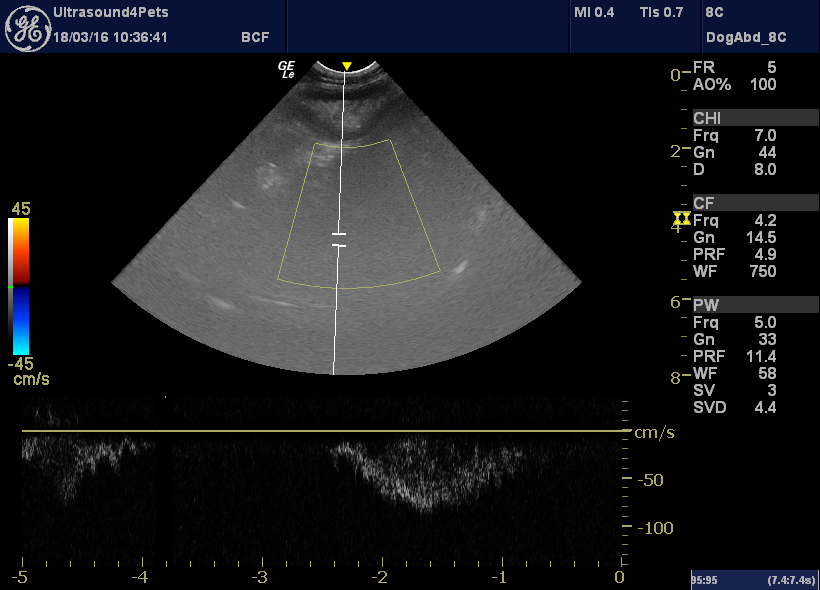
….and crystalluria….
And a third one, again a transverse view of the central area of the liver:
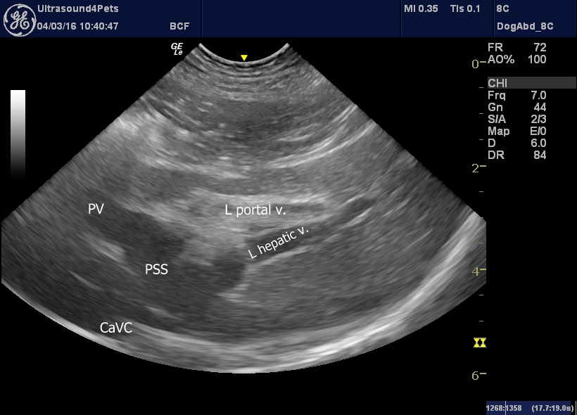
All three dogs had post-prandial bile acids >200.


