Some unusual bladder tumours in dogs
Transitional cell carcinomas are the commonest bladder neoplasm of dogs. However they only account for 70% of the total. Moreover, it’s important not to assume that all bladder tumours are TCCs since that may discourage partial cystectomy in situations where it may prove beneficial.
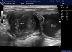
This lesion in an 8 year old dog was found on a survey of the abdomen requested when the owners ‘felt there was something wrong’! There were no urinary signs.

Sagittal plane view of the cranial pole of the bladder
The mass appears to be continuous with the muscular layer of the bladder wall which encouraged us to take him to surgery. Histopathological diagnosis was leiomyoma. Partial cystectomy was performed and the patient remains tumour-free at the time of writing.
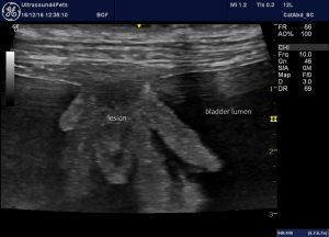
The second case remains unique in my experience. This dog presented with haematuria and frequency:

sagittal plane view of cranio-ventral bladder wall
Similarly, this is not a typical TCC. It does not involve the trigone and it is less heterogenous than most TCCs. However, there does appear to be obliteration of normal wall layering. Since the base was relatively narrow and cranially-sited in the bladder this lesion was also excised via partial cystectomy. Histopathological and immunohistochemical diagnosis…..
…….. T cell lymphoma!
Much discussion was devoted to deciding on post-op management of this case. Ultrasonography of abdominal and thoracic viscera was otherwise normal and a splenic core biopsy proved unremarkable.
Sadly, only a month later he presented again having deteriorated rapidly with anaemia, melaena and anorexia. This time there was multi-organ involvement:

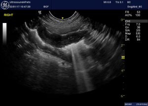
abnormal jejunum: there is wall thickening and obliteration of nomral layering

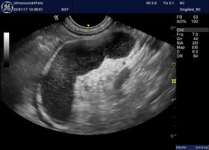
This jejunal lymph node is large, rounded and hypoechoic
Flow cytometry on samples harvested under ultrasound-guidance confirmed this to be T cell lymphoma


